
Method and system for extracting left atrial appendage data parameters based on CT images

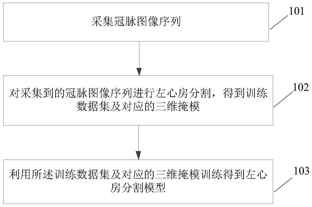
A CT image and parameter technology, applied in the field of image processing, can solve problems such as lack of friendliness of doctors who do not have the ability to analyze left atrial appendage parameters, deviation, and left atrial appendage closure surgery, and achieve the effect of facilitating clinical evaluation and scientific research analysis

Active Publication Date: 2021-09-07
CREALIFE MEDICAL TECH BEIJING
View PDF0 Cites 0 Cited by
  • Summary
  • Abstract
  • Description
  • Claims
  • Application Information

AI Technical Summary

Problems solved by technology

[0003] 1. Accurate measurement of the opening and dimension of the left atrial appendage requires clinicians to have strong imaging knowledge, and imaging specialists currently do not have the ability to analyze the parameters of the left atrial appendage. How to enable clinicians to obtain the required parameters based on clinical needs there are difficulties;
[0004] 2. At present, there is no application tool specifically for left atrial appendage analysis in clinical work. If image analysis functions of other applications are used to analyze left atrial appendage, there will be serious deviations. For beginners or doctors who have not been deeply involved in left atrial appendage closure surgery lack of friendliness

Method used


Image

  • Method and system for extracting left atrial appendage data parameters based on CT images
  • Method and system for extracting left atrial appendage data parameters based on CT images
  • Method and system for extracting left atrial appendage data parameters based on CT images

Examples


Embodiment Construction

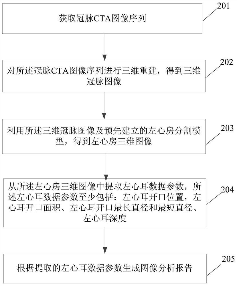
[0074] Aiming at the current clinical difficulties in applying CT images to the preoperative evaluation of the left atrial appendage, the present invention provides a method and system for extracting data parameters of the left atrial appendage based on CT images, and obtains three-dimensional coronary arteries by performing three-dimensional reconstruction on coronary CTA image sequences. Then use the 3D coronary image and the pre-established left atrium segmentation model to obtain the 3D image of the left atrium; extract the data parameters of the left atrial appendage from the 3D image of the left atrium, and obtain the 3D data set of the left atrial appendage, so as to provide comprehensive data for the closure of the left atrial appendage. , Reliable reference data are helpful for clinicians to quickly assess the risk of coronary heart disease in patients.

[0075] The left atrium segmentation model can be obtained by collecting a large number of coronary image sequence t...


PUM


Abstract

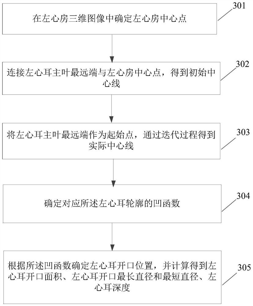
The present invention discloses a method and system for extracting data parameters of the left atrial appendage based on CT images. The method includes: acquiring a coronary CTA image sequence; performing three-dimensional reconstruction on the coronary CTA image sequence to obtain a three-dimensional coronary image; using the The three-dimensional coronary image and the pre-established left atrium segmentation model are used to obtain a three-dimensional image of the left atrium; the data parameters of the left atrial appendage are extracted from the three-dimensional image of the left atrium; the data parameters of the left atrial appendage at least include: the opening position of the left atrial appendage, the left atrial appendage Opening area, longest diameter and shortest diameter of LAA opening, LAA depth; generate an image analysis report based on extracted LAA data parameters. By using the present invention, various three-dimensional data of the left atrial appendage can be efficiently, quickly and accurately obtained, providing effective reference for subsequent clinical evaluation and scientific research analysis.

Description

technical field [0001] The invention relates to the field of image processing, in particular to a method and system for extracting left atrial appendage data parameters based on CT images. Background technique [0002] The left atrial appendage is an ear-shaped sac protruding from the left atrium, which is a part of the left atrium and is the main component of the left atrium. Left atrial appendage closure is currently a new trend in the treatment of stroke prevention in patients with atrial fibrillation around the world. It can effectively reduce the mortality and disability of patients, and reduce the occurrence of bleeding at the same time. Due to the complex anatomy of the left atrial appendage, in the prior art, doctors usually use CT imaging to evaluate the shape and variation of the left atrial appendage based on clinical experience, and perform related measurements to determine the anatomical structure of the left atrial appendage Related parameters provide data ref...

Claims


Application Information

Patent Timeline
Patent Type & Authority Patents(China)
IPC IPC(8): A61B6/03
CPCA61B6/032A61B6/5211A61B6/5217
Inventor 陈韬郭军陈韵岱王民汉周宇李晓光刘阁易永珍
Owner CREALIFE MEDICAL TECH BEIJING
Learn More