
Electronic endoscope system and data processing device

A technology of electronic endoscope and camera position, applied in image data processing, endoscope, rectal electron microscope, etc., can solve the problem of identifying lesion parts, which is not easy

Pending Publication Date: 2021-02-09
HOYA CORP
View PDF5 Cites 2 Cited by
  • Summary
  • Abstract
  • Description
  • Claims
  • Application Information

AI Technical Summary

Problems solved by technology

In addition, it is not easy for even skilled surgeons to identify the lesion from subtle differences in color, so careful work is required

Method used


Image

  • Electronic endoscope system and data processing device
  • Electronic endoscope system and data processing device
  • Electronic endoscope system and data processing device

Examples


Embodiment Construction

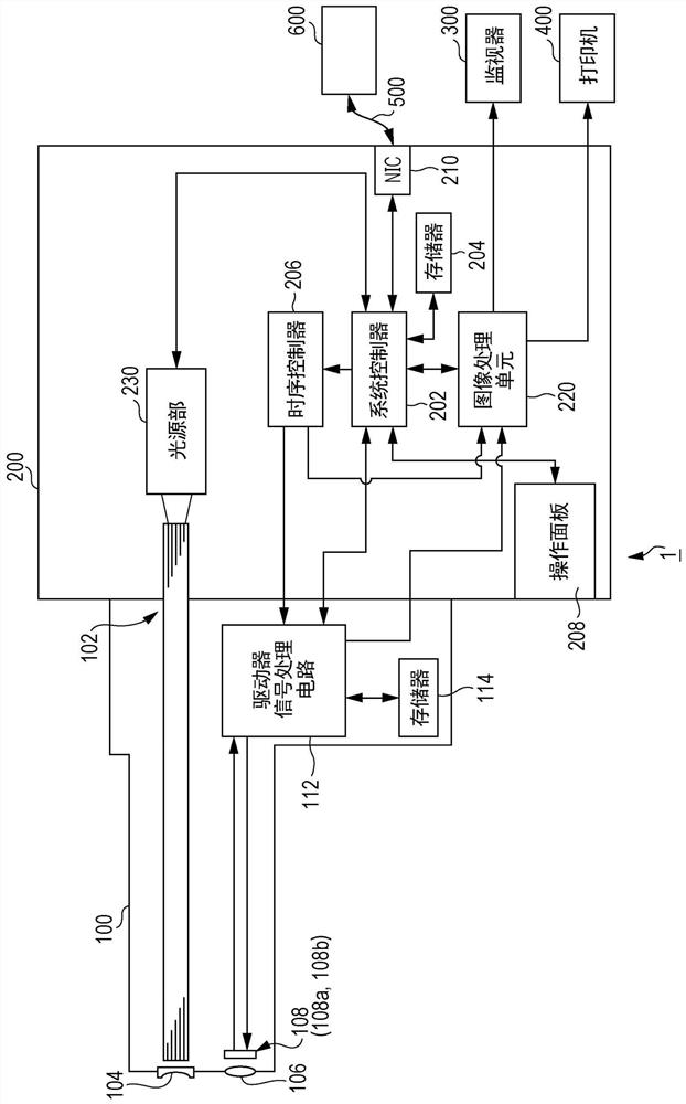
[0054] Hereinafter, before describing the electronic endoscope system and the data processing apparatus according to the embodiment of the present invention with reference to the drawings, first, a method for evaluating the spread of intra-organ lesions will be conceptually described.

[0055] (Outline of Assessment of Intra-Organ Lesion Degree)

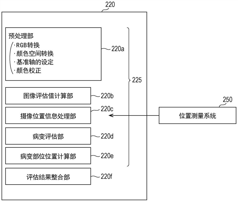
[0056] The processor of the electronic endoscope system in the embodiment described below processes the image of the living tissue in the organ captured by the electronic endoscope, and evaluates the degree of the lesion in the organ. The degree of lesions in the organ includes the spread of the lesions and the intensity of the lesions used to indicate the degree of progression of the lesions at each location, as factors affecting the degree of lesions in the organ. For example, when imaging living tissue inside an organ, an electronic scope is inserted from the open end of a tubular organ to the deepest position of the object to be ...


PUM


Abstract

An electronic endoscope system comprises an electronic endoscope for acquiring an image of biological tissue within an organ; a processor equipped with an evaluation unit for processing a plurality ofacquired images of the biological tissue to evaluate the extent of a lesion; and a monitor for displaying on a screen the evaluation result for the extent of the lesion. The evaluation unit comprises: an image evaluation value calculation unit whereby, for each of the plurality of images of the biological tissue, an image evaluation value is calculated, indicating the strength of the lesion in the biological tissue; and a lesion evaluation unit whereby, for each of a plurality of sections yielded by sectioning the organ interior region using information regarding the imaging position within the imaged organ, a representative evaluation value for the image evaluation value is calculated from the image evaluation values for the plurality of images corresponding to each of the plurality of sections, and whereby the spread of the lesion in the depth direction of the organ interior is evaluated using the representative evaluation value.

Description

technical field [0001] The present invention relates to an electronic endoscope system and a data processing device for evaluating the spread of lesions in living tissue in an organ. Background technique [0002] The lesions in the living tissue ranged from thinning, rough surface, red inflammation of the mucosal layer of the living tissue to ulcers with local defects in the mucosal layer and its underlying layers, and there were different grades of severity. For example, in the ulcer site where ulcerative colitis (UC: Ulcerative Colitis) occurs, it becomes whitish with white coating and pus-like mucus, while in the inflamed site, it becomes red with edema and hyperemia. Such lesions can be photographed and observed through an endoscopic system. [0003] However, in order for the surgeon to be able to identify the normal part and the diseased part by the color difference contained in the endoscopic image, a long training under the guidance of a skilled person is required. ...

Claims


Application Information

Patent Timeline
Patent Type & Authority Applications(China)
IPC IPC(8): A61B1/045A61B1/00
CPCA61B1/045A61B1/00006A61B1/05A61B1/0669A61B1/07A61B1/00045A61B1/00055A61B1/000094A61B1/00097G06T7/11A61B1/0005A61B1/31G06T7/60G06T2207/10068
Inventor 小泉辽平池本洋祐牧野贵雄
Owner HOYA CORP
Learn More