
Quantitative Analysis Method of Cerebral Vascular Morphological Characteristics

A morphological feature and quantitative analysis technology, applied in the field of medical imaging, can solve problems such as lack of quantitative methods and difficult tumor identification, and achieve the effect of promoting diagnosis and prevention research, promoting diagnosis and treatment research, and realizing early detection and treatment effect evaluation.

Active Publication Date: 2021-12-28
SHANGHAI UNITED IMAGING HEALTHCARE
View PDF0 Cites 0 Cited by
  • Summary
  • Abstract
  • Description
  • Claims
  • Application Information

AI Technical Summary

Problems solved by technology

The existing quantitative analysis methods of blood vessels can realize simple branch and radius calculation, but lack of other quantitative methods, it is difficult to be used for tumor identification

Method used


Image

  • Quantitative Analysis Method of Cerebral Vascular Morphological Characteristics
  • Quantitative Analysis Method of Cerebral Vascular Morphological Characteristics
  • Quantitative Analysis Method of Cerebral Vascular Morphological Characteristics

Examples


Embodiment Construction

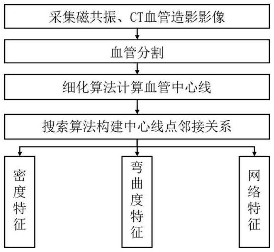
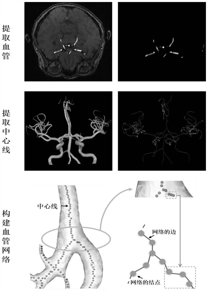
[0042] Hereinafter, taking the analysis of magnetic resonance cerebral vascular enhanced image (MRA-TOF) as an example, the specific embodiments of the present invention will be described in detail with reference to the accompanying drawings. figure 2 The flow chart of the MRA-TOF blood vessel analysis method provided by the present invention.

[0043] In step S1, a blood vessel image is extracted from the MRA-TOF, and the image only contains tubular structures.

[0044] In step S2, the vascular structure in step S1 is refined by using a morphological thinning algorithm, and the center line of the vascular structure is obtained, and the set of all points on the center line is denoted as C.

[0045] Step S3, judging the adjacency relationship between each point on the center line of the blood vessel, the distance between the two points is less than or equal to Then there is an adjacency relationship, and there is an adjacency relationship between points to form an edge, ther...


PUM


Abstract

The invention belongs to the technical field of medical imaging, and specifically relates to a quantitative analysis method for morphological characteristics of cerebral blood vessels. The invention utilizes the structural characteristics of the vascular network to realize the calculation of various quantitative features by constructing the adjacency matrix of the vascular network, has strong universality, and can be used for vascular images obtained by different acquisition methods such as X-ray, CT, and MRI. The analysis results include multiple characteristics such as the number of vascular branches, branch lengths, vascular radius, vascular tortuosity, and vascular network complexity, which can effectively improve the efficiency of doctors in analyzing quantitative parameters of blood vessels; It can realize the automatic analysis of two-dimensional blood vessel images and three-dimensional blood vessel images. The invention plays a huge role in the intelligent diagnosis and treatment of cardiovascular diseases and other major vascular diseases in the future, and has great market potential and economic and social benefits.

Description

technical field [0001] The invention belongs to the technical field of medical imaging, and particularly relates to a method for quantitative analysis of morphological features of cerebral blood vessels. Background technique [0002] The occurrence and development of many diseases are often accompanied by abnormalities and lesions of blood vessels in tissues and organs. With the rapid development of medical imaging technology, the use of various imaging methods to non-invasively observe tissue blood vessels, analyze and study the changes of blood vessels, play an important role in clinical practice. [0003] Vascular abnormalities and lesions are the main features of cardiovascular and cerebrovascular diseases, liver cirrhosis, various tumors and other diseases. For a long time, the method of quantitative analysis of tissue blood vessels is to use tissue slices to measure related parameters such as blood vessel density, blood vessel length and diameter. Such methods are in...

Claims


Application Information

Patent Timeline
Patent Type & Authority Patents(China)
IPC IPC(8): A61B5/02A61B5/055A61B5/00
CPCA61B5/02007A61B5/055A61B5/72A61B5/0042A61B2576/026
Inventor 王鹤张博宇
Owner SHANGHAI UNITED IMAGING HEALTHCARE
Learn More