Medical image analysis using machine learning and an anatomical vector

A medical image and vector technology, applied in the field of medical image analysis using machine learning and anatomical vectors, can solve problems such as large computing workloads

Pending Publication Date: 2021-08-20
BRAINLAB
View PDF0 Cites 0 Cited by
  • Summary
  • Abstract
  • Description
  • Claims
  • Application Information

AI Technical Summary

Problems solved by technology

This requires registration of patient images to atlas data, which involves a large computational effort

Method used


Image

  • Medical image analysis using machine learning and an anatomical vector
  • Medical image analysis using machine learning and an anatomical vector
  • Medical image analysis using machine learning and an anatomical vector

Examples


Embodiment Construction

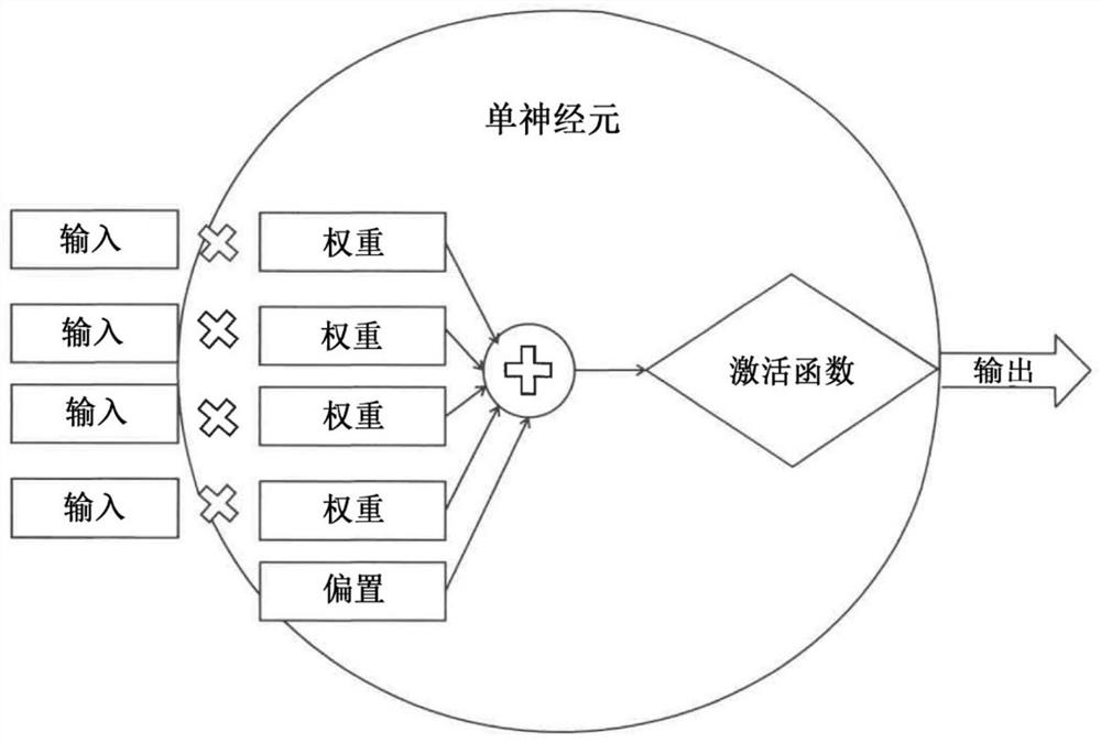
[0154] figure 1 Elucidate the structure of a neuron that is part of a neural network, such as a convolutional neural network, where inputs are assigned certain weights to be processed by an activation function that generates the neuron's output.

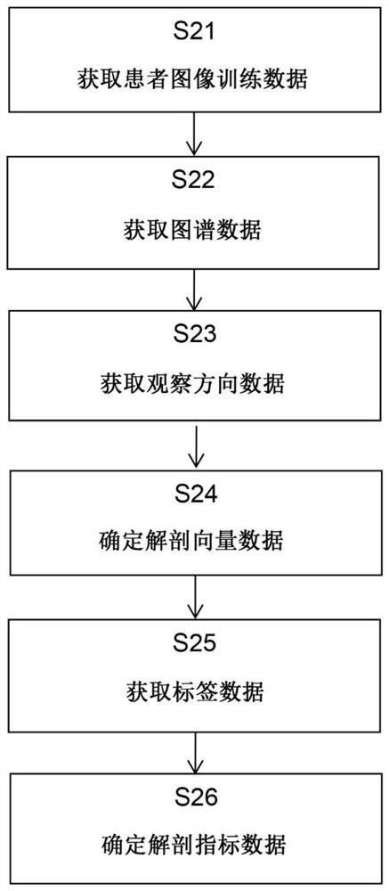
[0155] figure 2 The basic flow of the method according to the first aspect is described, starting from step S21, obtaining patient training image data, continuing to step S22, which includes obtaining atlas data, and then continuing to obtain viewing direction data in step S23. On this basis, step S24 calculates anatomical vector data, followed by acquiring label data in step S25. Finally, anatomical index data are determined in step S26.

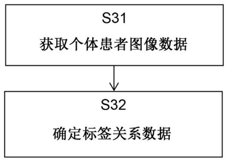
[0156] image 3 The basic steps of the method according to the second aspect are shown, wherein step S31 comprises acquiring individual patient image data and step 32 determines label relationship data.

[0157] Figure 4 The basic steps of the method according to the third aspect are shown...


PUM


Abstract

Disclosed is a computer-implemented method which encompasses registering a tracked imaging device such as a microscope having a known viewing direction and an atlas to a patient space so that a transformation can be established between the atlas space and the reference system for defining positions in images of an anatomical structure of the patient. Labels are associated with certain constituents of the images and are input into a learning algorithm such as a machine learning algorithm, for example a convolutional neural network,together with the medical images and an anatomical vector and for example also the atlas to train the learning algorithm for automatic segmentation of patient images generated with the tracked imaging device. The trained learning algorithm then allows for efficient segmentation and / or labelling of patient images without having to register the patient images to the atlas each time, thereby saving on computational effort.

Description

technical field [0001] The present invention relates to a computer-implemented method of training a learning algorithm to determine a relationship between, on the one hand, labels indicative of the location or type of anatomical structures in a medical image and, on the other hand, labels indicative of the location or type of anatomical structures in the medical image, As well as methods for segmenting and / or labeling medical patient images using trained learning algorithms, a corresponding computer program, a computer-readable storage medium storing such a program, a computer executing such a program, and a A system comprising an electronic data storage device and the aforementioned computer. Background technique [0002] Ability to use anatomical images to segment or label medical patient images. This requires registration of patient images to atlas data, which involves a large computational effort. Contents of the invention [0003] It is an object of the present inve...

Claims


Application Information

Patent Timeline
Patent Type & Authority Applications(China)
IPC IPC(8): G06T7/00G06T7/136G06K9/62G06N20/00
CPCG06T7/0012G06T7/136G06N20/00G06T2207/10061G06T2207/30004G06F18/214G06F18/24323G06V20/695G06V20/698G06V2201/03G06V10/82G06V10/764G06T7/0014G06T7/70G06V20/70G06V10/26G06V10/774G06T2207/10056G06T2207/20081G06T2207/20084
Inventor 斯特凡·维尔斯迈尔延斯·施马勒
Owner BRAINLAB
Why Eureka
Try Eureka