
AI-based image analysis for detecting normal images

A technology for normal and medical images, applied in image analysis, image enhancement, medical images, etc., can solve problems that cannot be solved and take up time for radiologists

Pending Publication Date: 2021-12-07
SIEMENS HEALTHCARE GMBH
View PDF8 Cites 0 Cited by
  • Summary
  • Abstract
  • Description
  • Claims
  • Application Information

AI Technical Summary

Problems solved by technology

Thus, while these machine learning algorithms may be helpful in general, they do not address the inherent problems of taking up radiologists' time and their broad applicability to a wide range of types of medical images and conditions

Method used


Image

  • AI-based image analysis for detecting normal images
  • AI-based image analysis for detecting normal images
  • AI-based image analysis for detecting normal images

Examples


Embodiment Construction

[0014] This disclosure is not limited to the particular systems, devices and methods described, as these may vary. The terminology used in this description is for the purpose of describing a particular version or embodiment only, and is not intended to limit the scope.

[0015] As used herein, the terms "algorithm", "system", "module" or "engine", if used herein, are not intended to limit the implementation and / or execution attributable thereto and / or performed thereby Any particular implementation of an action, step, process, etc. Algorithms, systems, modules, and / or engines may be, but are not limited to, software, hardware, and / or firmware, or any combination thereof, that perform specified functions, including, but not limited to, loading or storing in machine-readable memory in conjunction with processing Any use of a general-purpose and / or special-purpose processor with appropriate software executed by the processor. Additionally, unless otherwise specified, any names ...


PUM


Abstract

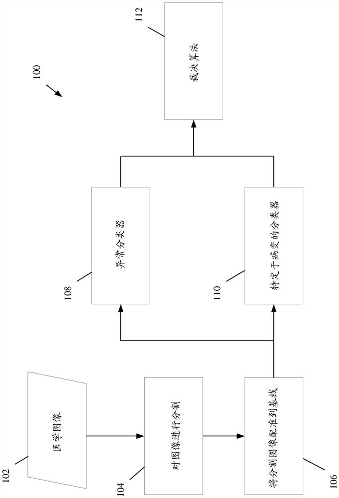
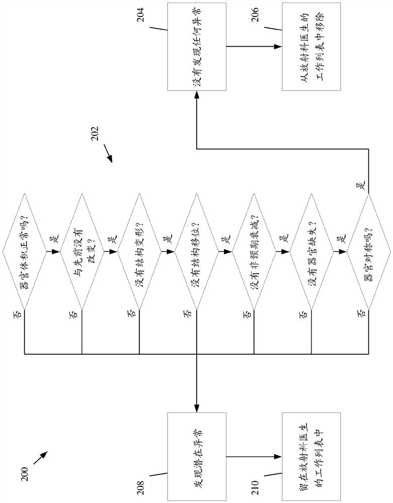
A system and method for identifying abnormal medical images are disclosed. The system can be configured to receive a medical image, segment an anatomical structure from the medical image to define a segmented dataset, register the segmented dataset to a baseline dataset defining a normal anatomical structure, classify, by an abnormality classifier, whether the anatomical structure within the medical image as either abnormal or normal, wherein the abnormality classifier comprises a machine learning algorithm trained to distinguish between normal and abnormal versions of the anatomical structure in medical images, and based on whether the anatomical structure can be segmented from the medical image, whether the segmented dataset can be registered to the baseline dataset, or a classification associated with the medical image output by the abnormality classifier, flagging the medical image as either normal or abnormal.

Description

[0001] Cross References to Related Applications [0002] This application claims priority to U.S. Provisional Patent Application No. 63 / 033,875, filed June 3, 2020, entitled "AI-BASED IMAGE ANALYSIS FOR THEDETECTION OF NORMAL IMAGES (ESPECIALLY FOR MULTI-INDICATION EXAMS)," which U.S. The Provisional Patent Application is hereby incorporated by reference in its entirety. Background technique [0003] The volume of medical images that need to be processed keeps growing every year, placing an increasing burden on the radiologists responsible for analyzing and interpreting these images. Additionally, due to routine medical practice, some types of medical images, such as chest x-rays or chest CT images (eg, for lung cancer screening) have a high rate of "normal" (ie, without any radiation Abnormal cases visible on photography). Reviewing what is normal can take up considerable radiologist time that could be better spent reviewing and analyzing medical images that actually have r...

Claims


Application Information

Patent Timeline
Patent Type & Authority Applications(China)
IPC IPC(8): G06T7/00G06T7/11G06T7/136G06K9/62G06N20/00
CPCG06T7/0012G06T7/11G06T7/136G06N20/00G06T2207/20081G06T2207/20084G06T2207/30004G06F18/241G06T2207/30096G16H40/20G16H30/20G16H30/40G06N5/04G06V2201/03G06F18/2433G06F18/2163
Inventor P·赫尔策R·弗兰克S·施密特J·斯佩尔
Owner SIEMENS HEALTHCARE GMBH
Learn More