Quantitative characterization method of cell membrane tension

A quantitative characterization and cell membrane technology, applied in the fields of nanoscience and biophysics, can solve the problem of not being able to obtain quantitative values ​​of cell membrane tension

Pending Publication Date: 2022-03-01
THE NAT CENT FOR NANOSCI & TECH NCNST OF CHINA
View PDF0 Cites 0 Cited by
  • Summary
  • Abstract
  • Description
  • Claims
  • Application Information

AI Technical Summary

Problems solved by technology

[0013] Characterization of cell membrane tension by measuring the tension of the tether using an atomic force microscope Although the magnitude of different cell membrane tensions can be qualitatively characterized, the quantitative value of the cell membrane tension cannot be obtained

Method used


Image

  • Quantitative characterization method of cell membrane tension
  • Quantitative characterization method of cell membrane tension
  • Quantitative characterization method of cell membrane tension

Examples


Embodiment 1

[0086] Example 1 Method for Characterizing Cell Membrane Tension Using Atomic Force Microscopy

[0087] 1. AFM indentation experiment and collection of force relaxation curve

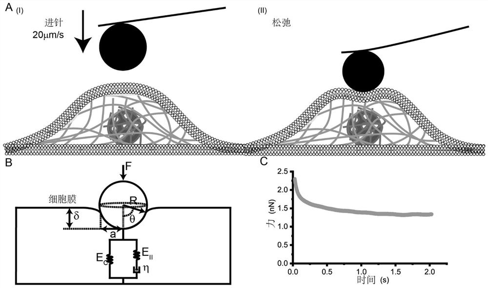
[0088] Such as image 3 As shown in middle A, taking the spherical probe as an example, the AFM probe tip approaches the cell surface at a rate of 20 μm / s to produce an indentation with a depth of about 1 μm, and then remains stationary for about 3 seconds until the force relaxation signal gradually stabilizes. Record distance, time and force data for the entire process. according to image 3 In the physical model shown in B, the analytical formula of the force relaxation process is derived theoretically. Then, combine the analytical expression with image 3 The force relaxation curve shown in C was fitted to obtain a quantitative solution of cell membrane tension.

[0089] 2. Analysis of AFM force relaxation curve considering membrane tension

[0090] Due to the effect of membrane tension, the pr...


PUM

PropertyMeasurementUnit
Diameteraaaaaaaaaa

Abstract

The invention provides a quantitative characterization method of cell membrane tension, which combines a cell membrane tension model and a cytoplasm viscoelasticity model, and performs quantitative characterization on the cell membrane tension by using an atomic force microscope. The method specifically comprises the following steps: (1) obtaining single cell force relaxation signal data based on an AFM nanoindentation experiment; (2) constructing a cell power relaxation curve equation; and (3) quantitative calculation of cell membrane tension. The detection means for quantitatively detecting the membrane tension of the living cells, provided by the invention, is helpful for people to have further deeper understanding on cell behaviors, and also provides a new view angle and direction for people to carry out early diagnosis, regulation and treatment on various diseases by regulating the physical characteristics of the cells.

Description

technical field [0001] The invention relates to nanometer science and biophysics, in particular to a quantitative characterization method of cell membrane tension. Background technique [0002] Existing techniques commonly used to characterize the mechanical properties of cell membranes mainly include micropipettes and atomic force microscopy. Among them, the micropipette is mainly aimed at suspension cells, and the characterization of cell membrane tension can be realized through the Laplace formula. The atomic force microscope is mainly aimed at adherent cells. It can compare the tension of different cell membranes according to the tension of the tether on the surface of the cell membrane, but it cannot realize the quantitative detection of the tension of the cell membrane. In addition, atomic force microscopy is also used to characterize the viscoelasticity of cells, but the role of cell membranes is often ignored in this process. [0003] ①Measurement of cell membrane ...

Claims


Application Information

Patent Timeline
IPC IPC(8): G01N15/00
CPCG01N15/00G01N15/01
Inventor 任克礼韩东
Owner THE NAT CENT FOR NANOSCI & TECH NCNST OF CHINA
Why Eureka
Try Eureka