
Brain image-based hemorrhagic area determination method and device, equipment and medium

A region-determining, region-based technology, applied in the field of hemorrhage region determination based on brain images, can solve problems such as diagnostic errors, subjective factors, and low recognition rate of hemorrhage regions, and achieve high accuracy and fast recognition speed

Pending Publication Date: 2022-04-08
沈阳东软智能医疗科技研究院有限公司
View PDF0 Cites 2 Cited by
  • Summary
  • Abstract
  • Description
  • Claims
  • Application Information

AI Technical Summary

Problems solved by technology

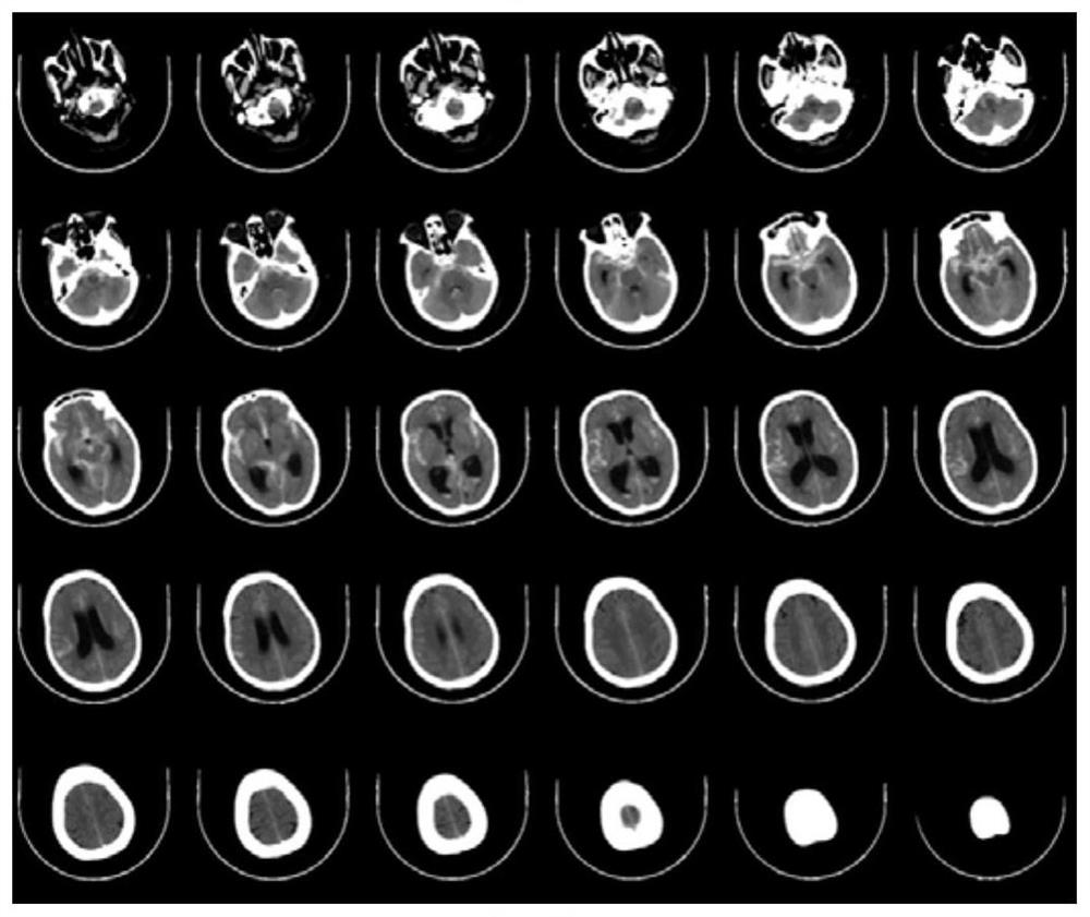
[0004] At present, determining whether the hemorrhage area invades the ventricle is basically achieved by observing the brain CT with human eyes, which is not only affected by subjective factors, but also has a low recognition rate when the hemorrhage area is not clearly developed, which will lead to large diagnostic errors

Method used


Image

  • Brain image-based hemorrhagic area determination method and device, equipment and medium
  • Brain image-based hemorrhagic area determination method and device, equipment and medium
  • Brain image-based hemorrhagic area determination method and device, equipment and medium

Examples


Embodiment Construction

[0047] The following will clearly and completely describe the technical solutions in the embodiments of the application with reference to the drawings in the embodiments of the application. Apparently, the described embodiments are only some of the embodiments of the application, not all of them. All other embodiments obtained by persons of ordinary skill in the art based on the embodiments in the present application without creative efforts shall fall within the protection scope of the present application.

[0048] It should be noted that the terms "first" and "second" in the description and claims of the present application and the above drawings are used to distinguish similar objects, but not necessarily used to describe a specific sequence or sequence. It is to be understood that the data so used are interchangeable under appropriate circumstances such that the embodiments of the application described herein can be practiced in sequences other than those illustrated or des...


PUM


Abstract

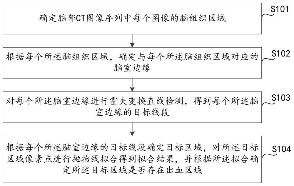
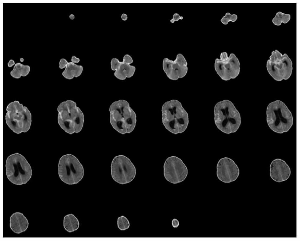
The invention provides a method, a device and equipment for determining a bleeding area based on a brain image, and a medium. The method comprises the following steps: determining a brain tissue area of each image in a brain CT image sequence; according to each brain tissue area, determining a ventricular edge corresponding to each brain tissue area; performing Hough transform straight line detection on each ventricular edge to obtain a target line segment of each ventricular edge; and determining a target area according to the target line segment of each ventricle edge, performing parabola fitting on pixel points of the target area to obtain a fitting result, and determining whether a bleeding area exists in the target area or not according to the fitting result. According to the method, the bleeding area is automatically determined based on the brain CT image, manual participation is not needed, the recognition speed is high, and the accuracy rate is high.

Description

technical field [0001] The embodiments of the present application relate to image processing technologies, and in particular to a method, device, device and medium for determining a hemorrhage area based on brain images. Background technique [0002] Intracerebral hemorrhage (ICH) is a type of stroke, accounting for about 13% of strokes. It is caused by rupture of cerebral arteries and causes localized bleeding in surrounding tissues. Intracerebral hemorrhage includes traumatic hemorrhage and primary cerebral hemorrhage. Cerebral hemorrhage has the characteristics of acute onset, critical and complex conditions, high mortality and disability rates, and has become the main cause of adult disability and death, seriously endangering the health of the people. [0003] Whether the hemorrhage area invades the ventricle is an important index to measure the severity of cerebral hemorrhage. The ventricle is a pathway through which the cerebrospinal fluid circulates in the human body...

Claims


Application Information

Patent Timeline
Patent Type & Authority Applications(China)
IPC IPC(8): G06T7/00G06T7/11G06T7/136G06V10/48G06V10/762G06V10/764G06K9/62
Inventor 马丽娟张霞蔡巍
Owner 沈阳东软智能医疗科技研究院有限公司
Learn More