System, device and method for photographing and checking anterior segment and posterior segment of eye by mobile phone

A mobile phone and eye technology, applied in the field of medical devices, can solve problems such as large differences in the position and size of the camera, unsatisfactory use effect of the connecting bracket, and shaking of the slit lamp microscope lens, etc., to achieve stable photographing process, convenient eye inspection work, fast The effect of taking pictures to check

Pending Publication Date: 2022-06-07
中国人民解放军联勤保障部队第九〇〇医院
View PDF0 Cites 0 Cited by
  • Summary
  • Abstract
  • Description
  • Claims
  • Application Information

AI Technical Summary

Problems solved by technology

[0003] When using a slit lamp microscope, in order to facilitate the inspection and ensure the inspection effect, the mobile phone is often fixed on the slit lamp microscope lens barrel with a connecting bracket, and the camera of the mobile phone is aligned with the slit lamp microscope lens barrel to take pictures of the patient's eyes, but due to different mobile phones The position and size of the camera vary greatly, and the effect of using various connecting brackets is not ideal, and you need to tap the screen when taking pictures, which will easily shake the microscope lens of the slit lamp and make the operation inconvenient

Method used


Image

  • System, device and method for photographing and checking anterior segment and posterior segment of eye by mobile phone
  • System, device and method for photographing and checking anterior segment and posterior segment of eye by mobile phone
  • System, device and method for photographing and checking anterior segment and posterior segment of eye by mobile phone

Examples


Embodiment 1

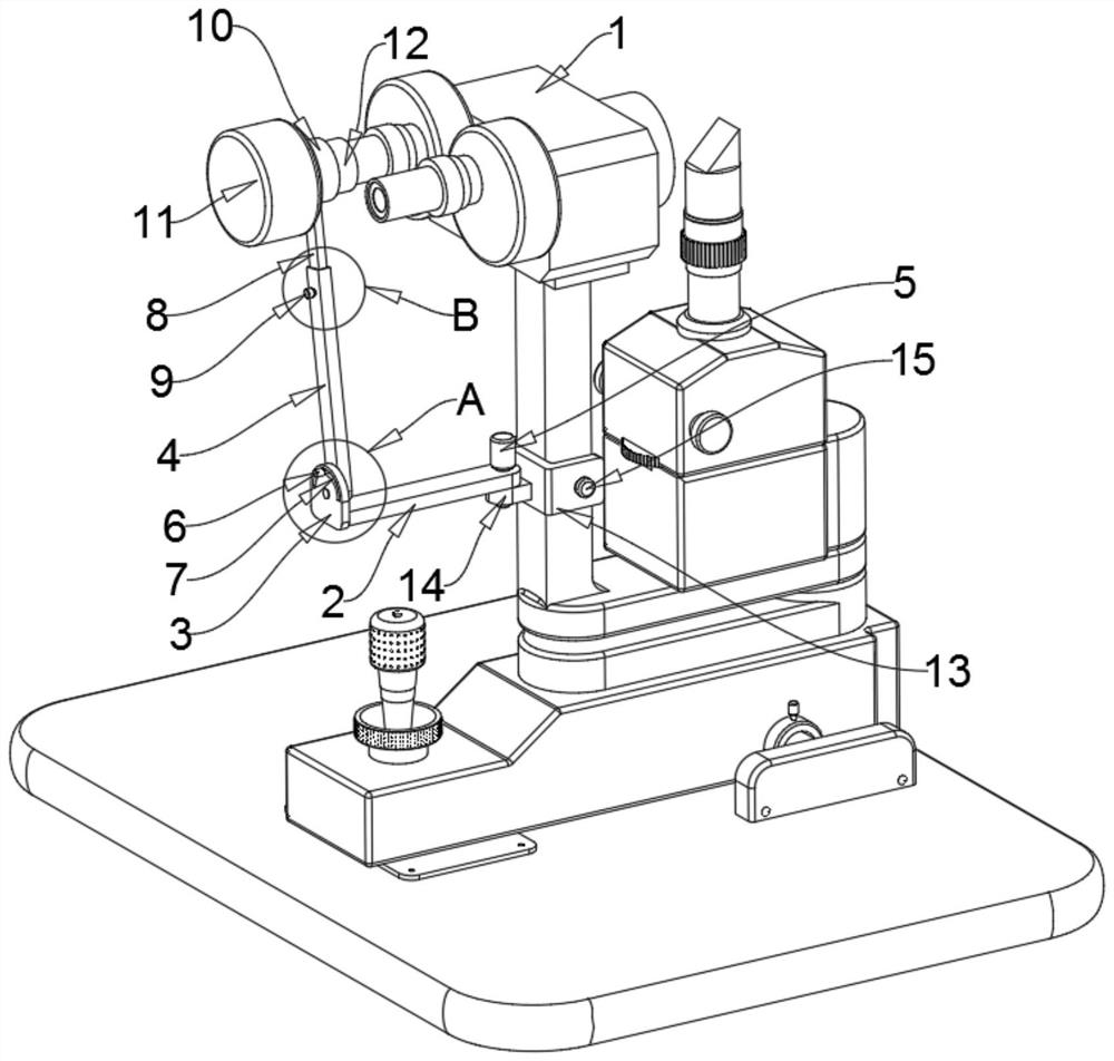
[0043] A device for realizing photographing inspection of the anterior and posterior segments of the eye by a mobile phone, comprising a mobile terminal, a camera module 11 and a slit lamp microscope 1, wherein the camera module includes a second wireless signal transceiver unit, a second processor and a camera unit , the second signal transceiver unit is bidirectionally connected with the second processor, the second processor is bidirectionally connected with the camera unit, the lens support arm of the slit lamp microscope 1 is provided with a detachable fixing mechanism, and the fixing mechanism is rotatably connected to support the camera module 11, the mobile terminal is also equipped with an auxiliary inspection device for auxiliary inspection of eyes.

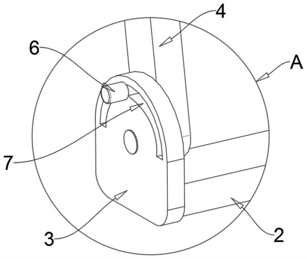
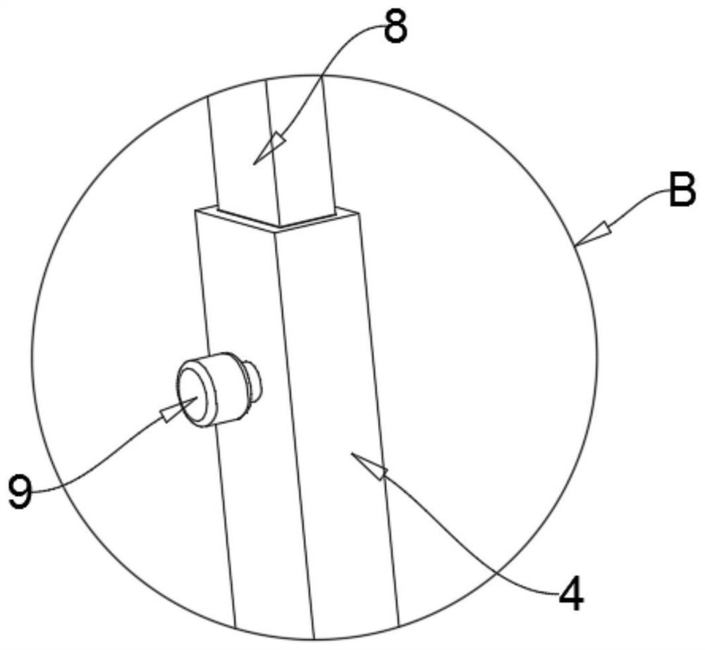
[0044] The support mechanism includes a primary rotating rod 2, a connecting plate 3, a secondary rotating rod 4, a primary threaded knob 5 and a secondary threaded knob 6. The primary threaded knob 5 is vertically rotat...

Embodiment 2

[0049] A device for realizing photographing inspection of anterior and posterior segments of an eye with a mobile phone, on the basis of Embodiment 1, the mobile terminal includes a camera, a signal input unit, a first processor, an image processing unit, and a first wireless signal transceiver unit, The camera is electrically connected with the first processor, the signal input unit and the image processing unit are both bidirectionally connected with the first processor, and the first processor is bidirectionally connected with the first wireless signal transceiving unit.

[0050] Specifically, the first wireless signal transceiver unit and the second wireless signal transceiver unit are connected through a wireless network, and the mobile terminal is a mobile phone.

Embodiment 3

[0052] A device for realizing photographing inspection of the front and rear segments of the eye with a mobile phone, on the basis of the second embodiment, the fixing mechanism includes a U-shaped frame 13, an inner threaded ear plate 14, a four-level threaded knob 15, and a first-level gasket 16. and the secondary washer 17, the primary washer 16 is fixedly connected to the inner side of the opening of the U-shaped frame 13, and the fourth-stage threaded knob 15 is threadedly connected to the side of the U-shaped frame 13 away from the primary washer 16 and penetrates the U-shaped frame. 13. The secondary washer 17 is fixedly connected to the end of the fourth-level threaded knob 15, the primary washer 16 and the secondary washer 17 are respectively pressed against both sides of the support arm of the lens of the slit lamp microscope 1, and the inner threaded ear plate 14 is horizontally fixed Connected to the outside of the U-shaped frame 13 , the primary threaded knob 5 is ...


PUM


Abstract

The invention discloses a system, a device and a method for photographing and checking anterior and posterior segments of eyes by a mobile phone, and relates to the field of medical instruments. According to the scheme, the system comprises a mobile terminal, a camera shooting module and a slit-lamp microscope, the camera shooting module comprises a second wireless signal receiving and transmitting unit, a second processor and a camera shooting unit, a detachable fixing mechanism is arranged on a lens supporting arm of the slit-lamp microscope, and a supporting mechanism used for supporting the camera shooting module is rotationally connected to the fixing mechanism. The beneficial effects of the invention are that the photographing system used in the invention realizes the photographing inspection of the anterior segment and posterior segment of the eye through the cooperation of the mobile phone and the camera module, and realizes the versatility which is not possessed by the cooperation of a traditional connecting support and the mobile phone for photographing; the fixing mechanism and the supporting mechanism are arranged to fix and support the camera module, the camera module is controlled by the mobile phone to photograph, the slit-lamp microscope does not need to be touched in the photographing process, and the photographing effect is further improved; in addition, when no slit-lamp microscope exists, the anterior segment and posterior segment of the eyes of the patient can be conveniently and quickly photographed and examined by matching the mobile phone terminal with the auxiliary examination device.

Description

technical field [0001] The invention relates to the field of medical devices, in particular to a system, a device and a method for realizing photographing inspection of the anterior and posterior segments of the eye with a mobile phone. Background technique [0002] Slit lamp microscope is an essential and important instrument for ophthalmic examination. The slit lamp microscope is composed of an illumination system and a binocular microscope. It can not only observe the superficial lesions very clearly, but also adjust the focus and the width of the light source to make an "optical section", so that the lesions in the deep tissue can also be clearly displayed. . [0003] When using the slit lamp microscope, in order to facilitate the inspection and ensure the inspection effect, the mobile phone is usually fixed on the slit lamp microscope barrel with a connecting bracket, and the camera of the mobile phone is aligned with the slit lamp microscope barrel to take pictures of...

Claims


Application Information

Patent Timeline
Patent Type & Authority Applications(China)
IPC IPC(8): A61B3/135A61B3/00A61B3/14
CPCA61B3/135A61B3/14A61B3/0016A61B3/0075
Inventor 严伟明陈艳津陈小红叶倩江川夏雨昕王雨彤陈镁林星垂洪博发王云鹏陈梅珠
Owner 中国人民解放军联勤保障部队第九〇〇医院
Why Eureka
Try Eureka