Method and device for detecting thin cap fibroplaque based on intracranial artery image

A fiber and image technology, applied in the field of thin fiber cap plaque detection, can solve the problems of limited OCT image resolution, interference tasks, and difficulty in achieving precise segmentation with small errors, so as to achieve accurate and controllable segmentation process and improve accuracy , Improve the effect of model segmentation performance

Active Publication Date: 2022-08-09
UNIVERSITY OF CHINESE ACADEMY OF SCIENCES +2
View PDF12 Cites 0 Cited by
  • Summary
  • Abstract
  • Description
  • Claims
  • Application Information

AI Technical Summary

Problems solved by technology

Plaque boundaries in OCT images are usually not obvious. Relying on existing segmentation techniques, it is difficult to achieve accurate segmentation with small errors. Larger segmentation errors are unavoidable and will interfere with subsequent tasks.
On the other hand, due to the limited resolution of OCT images, segmentation errors cause huge interference to TCFA detection

Method used


Image

  • Method and device for detecting thin cap fibroplaque based on intracranial artery image
  • Method and device for detecting thin cap fibroplaque based on intracranial artery image
  • Method and device for detecting thin cap fibroplaque based on intracranial artery image

Examples


Embodiment Construction

[0071] The specific embodiments of the present invention will be further described below with reference to the accompanying drawings. The following examples are only used to illustrate the technical solutions of the present invention more clearly, and cannot be used to limit the protection scope of the present invention.

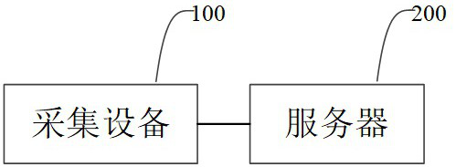
[0072] The method for detecting thin fibrous cap plaques based on intracranial arterial images provided by the embodiments of the present invention can be applied to, for example, figure 1 In the system architecture shown, the system architecture includes a collection device 100 and a server 200 .

[0073] Specifically, the acquisition device 100 is used to acquire an intracranial artery OCT image.

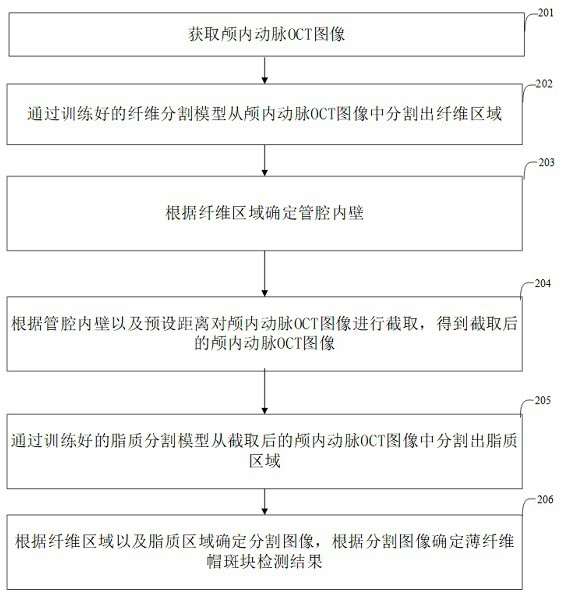
[0074] The server 200 is configured to segment the fiber region from the intracranial artery OCT image by using the trained fiber segmentation model; determine the lumen inner wall according to the fiber region; and intercept the intracranial artery OCT image a...


PUM


Abstract

The invention discloses a thin fibrous cap plaque detection method and device based on an intracranial artery image. The method comprises the following steps: acquiring an intracranial artery OCT image; segmenting a fiber region from the intracranial artery OCT image through the trained fiber segmentation model; determining the inner wall of the lumen according to the fiber area; intercepting the intracranial artery OCT image according to the inner wall of the lumen and a preset distance to obtain an intercepted intracranial artery OCT image; segmenting a lipid region from the intercepted intracranial artery OCT image through a trained lipid segmentation model; and determining a thin cap fibroplaque detection result according to the segmented image. According to the method, the characteristic difference between fibers and lipids is fully considered, two-section segmentation is adopted, namely, the fiber region is segmented through the trained fiber segmentation model, and then the lipid region is segmented through the trained lipid segmentation model, so that the model segmentation performance is improved, the segmentation process is more accurate and controllable, and the segmentation efficiency is improved. And the accuracy of the detection result of the thin fiber cap plaque is improved.

Description

technical field [0001] The invention relates to the field of computer technology, in particular to a method and a device for detecting thin fiber cap plaques based on intracranial arterial images. Background technique [0002] Thin-Cap Fibroatheroma (TCFA) is one of the characteristics of vulnerable plaques. Since such lesions have extremely high requirements for the thickness of the fibrous cap, no related reports have been reported in intracranial arteries. With the gradual application of optical coherence tomography (OCT) to intracranial arteries, people have a detectable method for intracranial arterial TCFA. [0003] Existing technologies focus on the intelligent interpretation of cardiovascular OCT images, and there is little work to explore and study intracranial arterial OCT. At present, no studies have clearly pointed out that the image interpretation and pathological treatment of cardiovascular OCT are completely applicable to intracranial arteries, and there is ...

Claims


Application Information

Patent Timeline
IPC IPC(8): G06T7/00G06T7/11G06T7/13G06T7/136G06T7/60
CPCG06T7/0012G06T7/11G06T7/13G06T7/136G06T7/60G06T2207/10101G06T2207/20016G06T2207/20081G06T2207/30101
Inventor 钟丽群焦力群杨戈徐然王韬罗沁轩冯瑶马妍杨斌
Owner UNIVERSITY OF CHINESE ACADEMY OF SCIENCES
Why Eureka
Try Eureka