Method and arrangement for determining an object contour

a technology of object contour and method, applied in the field of method and arrangement for determining object contour, can solve the problems of sensitive object contour obtained, large computational burden, and inability to administrate all images, and achieve the effect of reducing the complexity of image processing

Active Publication Date: 2008-11-11
CELLAVISION
View PDF6 Cites 33 Cited by
  • Summary
  • Abstract
  • Description
  • Claims
  • Application Information

AI Technical Summary

Benefits of technology

[0027]Such a method has been found to result in a quickly computed approximative GVF field, denoted AGVF field below. The computation of the AGVF field is performed so that, although the resulting AGVF field differs somewhat from the traditional GVF field, the segmentation results for white blood cells appear to be mainly unaffected. Using a method according to the invention, the time for using snakes with GVF performance in the segmentation can thus be considerably lowered. The present invention therefore makes it possible to use an improved segmentation in commercially available automatical scanning microscopes for analysis of white blood cells.

Problems solved by technology

An apparent drawback of binary thresholding is that the object contour that is obtained is sensitive to the level of the chosen threshold intensity, for example when the illumination conditions vary within av cell image.
Since the contour may have arbitrary shapes, it is relatively complicated and computationally heavy to administrate all the image elements of the contour, their neighbours and their matual relative positions.
Since the snake is represented by a limited number of parameters, its possible shapes are limited.
For white blood cells, which have fairly regular contours, this limitation is seldom a drawback.
Such a start may be easy for a human operator but harder to accomplish using automatic image analysis.
The greatest drawback of such a vector force field is that the amount of force rapidly descends as the coordinates (x,y) move away from those points that have an edge image f(x,y) that differs from zero.
Thus, such a vector force field does not work very well when the snake is started from a distance relatively far away from the sought object contour.
One previously known way of improving the ability of the vector force field to act at a distance is to smear the edge image, before or after the calculations of differences with for example a two-dimensional Gaussian filter, but that may result in other problems like details that become less apparent.
In the thesis it is pointed out that the timing performance of a GVF-snake, which uses this fast way of iterating the parameters, will be limited by the time it takes to calculate the GVF-field.
For an analysis of 200 white blood cells that corresponds to a total segmentation time of close on one minute, which limits the performance of existing automatic brightfield microscopes unless extra, expensive hardware is added.

Method used


Image

  • Method and arrangement for determining an object contour
  • Method and arrangement for determining an object contour
  • Method and arrangement for determining an object contour

Examples


Embodiment Construction

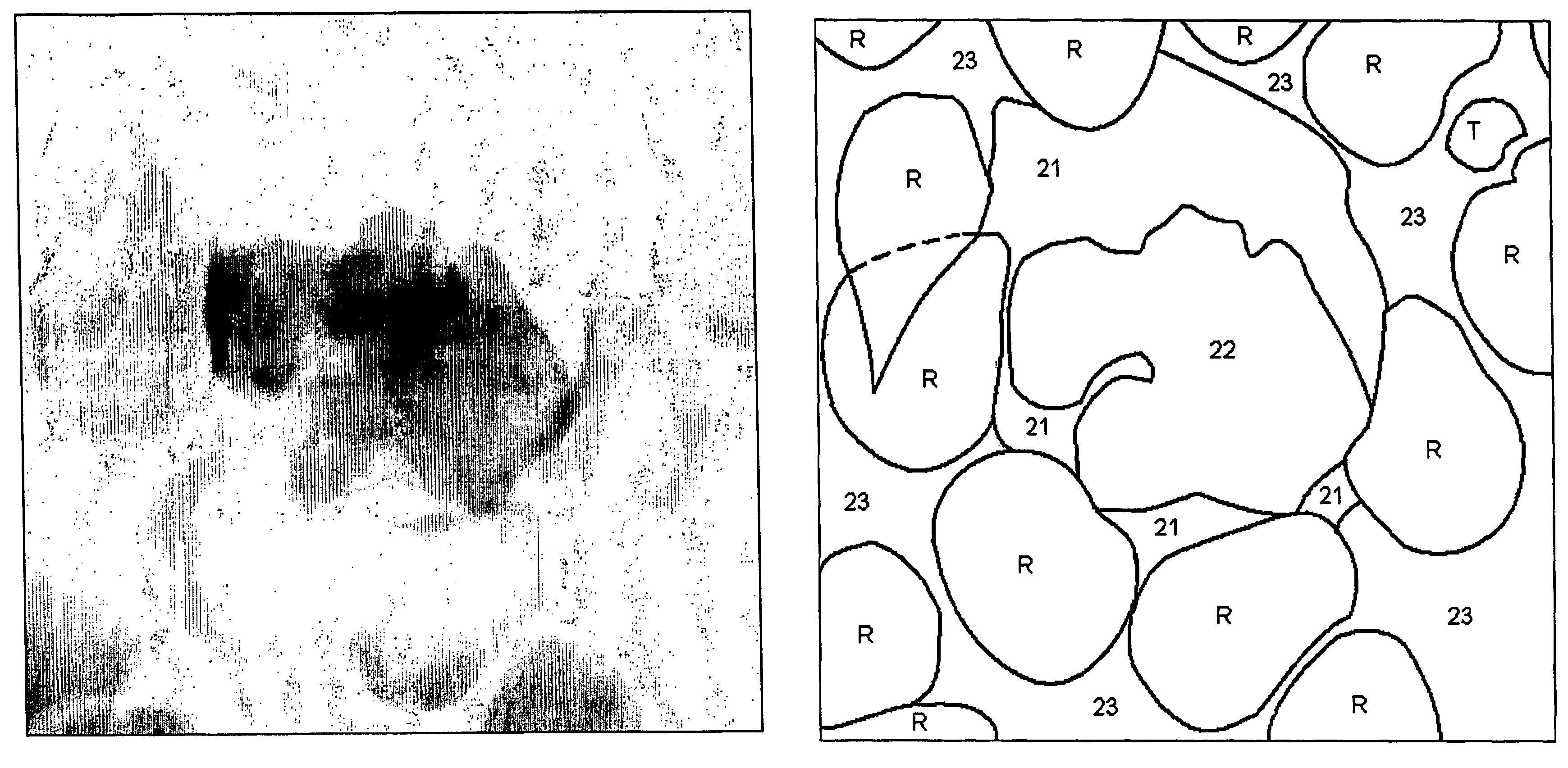
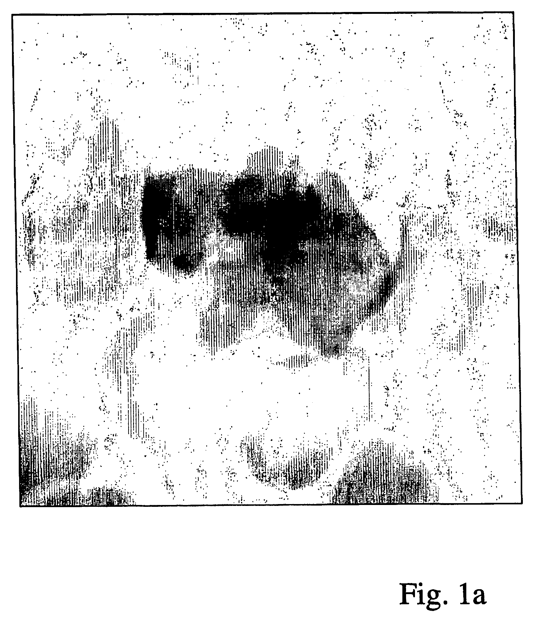
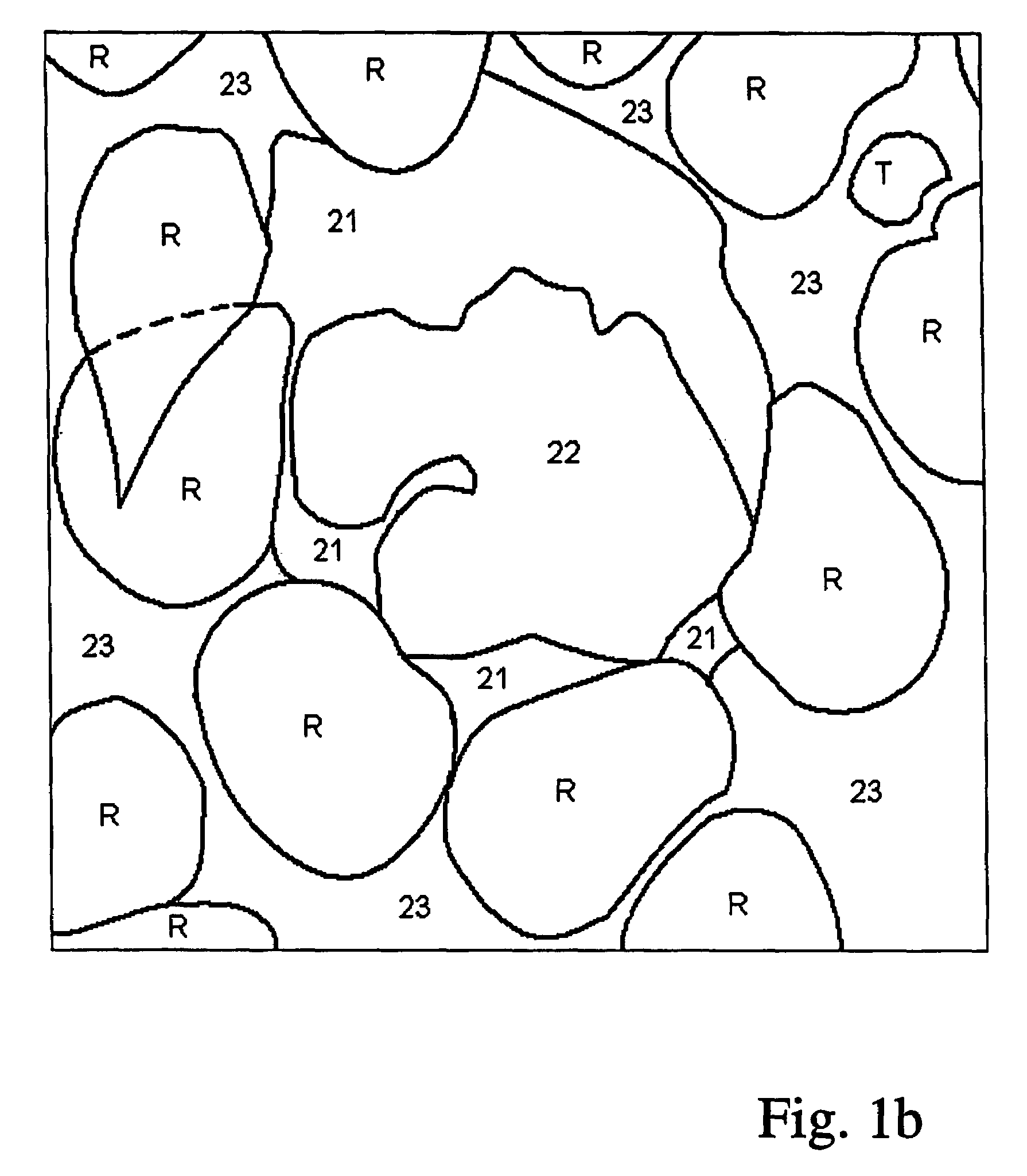
[0042]A white blood cell consists, from a segmentation point of view, of two parts—the cell nucleus 22 and the surrounding cytoplasm 21. The results of segmentation of the two parts are to some amount dependent upon one another:

[0043]In order to succeed in finding the border between cytoplasm and background using automatic image analysis in spite of the presence of adjacent cells, marked R and T in FIG. 1b, it is useful to be able to start the snake from a so called seed contour which is completely inside the cell. Therefore one wishes to have access to an estimate, for example a segmentation, of the cell nucleus as a so called seed contour for the snake.

[0044]In order to simplify the segmentation of the cell nucleus it is, on the other hand, good to have access to an image where there is only cytoplasm and cell nucleus left—i.e. an image where the cell already is segmented from the background and adjacent cells. To avoid an iterative process, a preliminary segmentation, see FIG. 2,...


PUM


Abstract

A method is disclosed for determining a sought object contour in a digital microscope image, which includes a plurality of image elements and reproduces a biological material. The method includes assigning edge values to at least a first subset of the image elements in the image; assigning values of a first gradient vector component whose values each includes a first linear combination of edge values of some surrounding image elements to at least a second subset of the image elements in the image; assigning values of a second gradient vector component whose values each include a second linear combination of edge values of some surrounding image elements to at least a third subset of the image elements in the image; and calculating an estimate of the sought object contour based upon values of the first and second gradient vector components.

Description

[0001]Cross reference to related application: This application claims benefit from Swedish patent application no SE-0302114-4, filed Jul. 21, 2003.FIELD OF THE INVENTION[0002]The present invention relates to a method of determining a sought object contour in a digital microscope image according to the preamble of claim 1. The invention also concerns a corresponding arrangement according to the preamble of claim 5 and a digital storage medium comprising a corresponding computer program according to the preamble of claim 6.BACKGROUND ART[0003]The blood of humans contains red blood cells, platelets and white blood cells. In medical applications there is often an interest in classifying the white blood cells into approximately twenty different sub classes and in estimating the distribution of these subclasses for individual patients. Traditionally this classification may be performed in a laborious way by a human expert looking through the eyepiece of a brightfield microscope and thereb...

Claims


Application Information

Patent Timeline
Patent Type & Authority Patents(United States)
IPC IPC(8): G06K9/48G06K9/00G06TG06T5/00
CPCG06K9/00127G06K9/0014G06T7/0083G06T2207/10056G06T2207/20056G06T2207/30024G06T7/12G06V20/69G06V20/695
Inventor MORELL, ADAM
Owner CELLAVISION
Why Eureka
Try Eureka