
Immune cell microfluid array

An immune cell and microfluidic technology, which is applied in the determination/inspection of microorganisms, material inspection products, measuring devices, etc., can solve the problems of inability to culture, inability to further observe cell growth status, and low inspection efficiency

Inactive Publication Date: 2004-08-11
中国人民解放军南京军区联勤部军事医学研究所
View PDF0 Cites 0 Cited by
  • Summary
  • Abstract
  • Description
  • Claims
  • Application Information

AI Technical Summary

Problems solved by technology

The main problem that this prior art exists is that the first is that the detection efficiency is low, because the confirmed leukocyte differentiation antigen has 166 molecular groups and a large number of subgroups, and it is very cumbersome to test one by one; Therefore, it is impossible to further observe the growth status of cells with different leukocyte differentiation antigens

Method used


Image

  • Immune cell microfluid array
  • Immune cell microfluid array

Examples


Embodiment 1

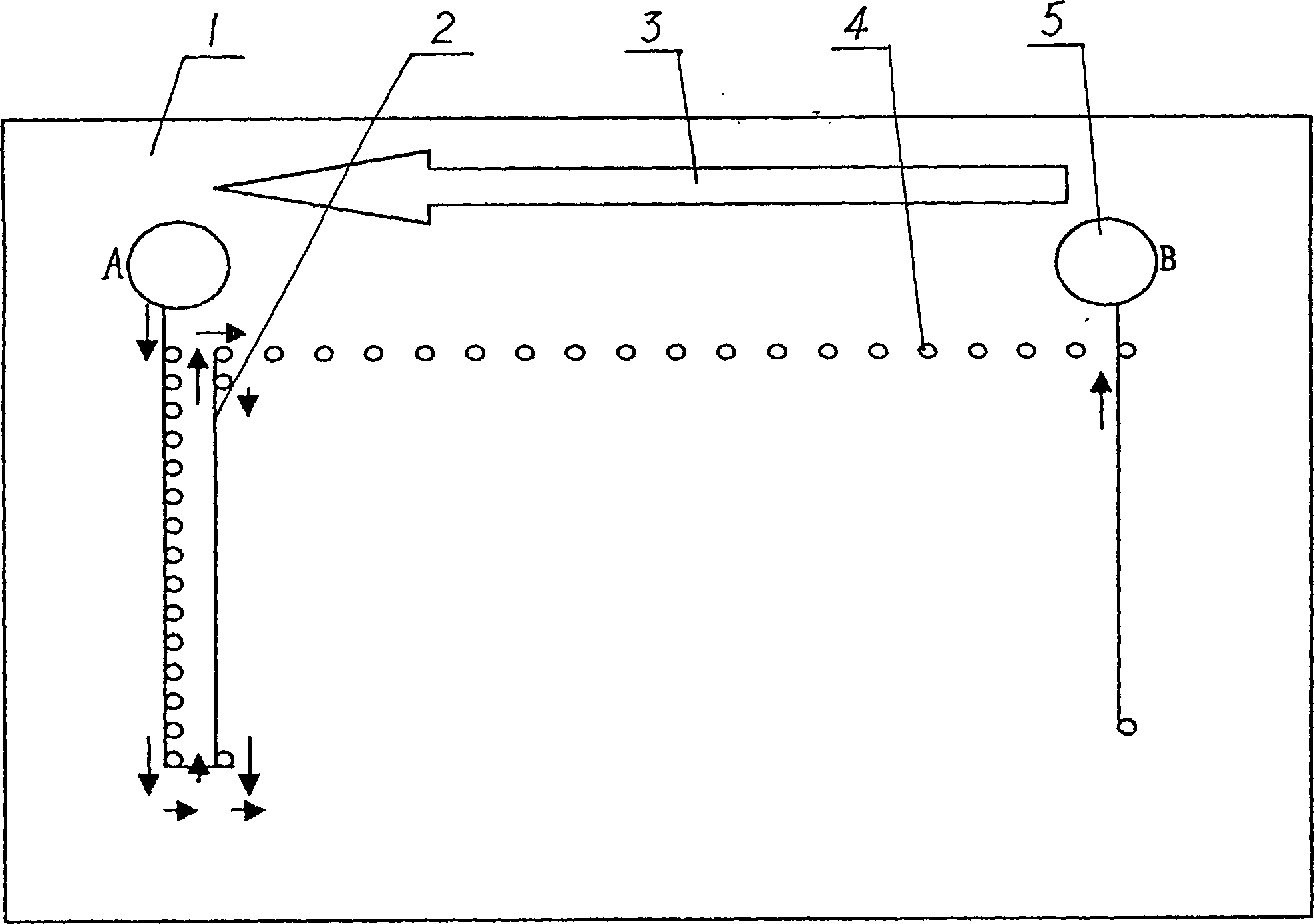
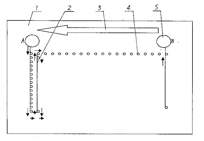
[0012] The immune cell microfluidic array of this embodiment is as figure 1 As shown, a 15×20 (or 10×20) immune (cell) microarray well 4 is prepared on a non-cytotoxic polymer compound bottom plate 1 . The thickness of the bottom plate 1 is 7-10mm, the diameter of each counterbore 4 is 2mm±0.2mm, the distance between adjacent counterbores is 2-3mm, and the micro-pipes 2 with a diameter of 0.1-0.5mm communicate with each other. The connected micro-pipes of two adjacent counterbores are obliquely placed according to the following rules, the opening of the micro-pipes sorted on the front hole is 0.8-1.0 mm away from the bottom of the hole, and the opening of the micro-pipes sorted on the back hole is 0.1-0.25 mm away from the bottom of the hole; By analogy, continue to form a reciprocating S-shaped circulation flow channel, so that all counterbores are connected. Experiments show that the above design parameters are suitable for cell immunoassay.

[0013] At both ends of the mi...

Embodiment 2

[0017] The basic situation of this embodiment is the same as that of the first embodiment, the difference is that the whole process is operated in a sterile environment because of the need for further research on cell growth and reproduction. In order to ensure the aseptic effect of the later cultivation, the polymer compound bottom plate is equipped with an isolation cover. After completing the same operation as in Example 1, put the lid on and put it into the incubator, and periodically replace the culture agent through the cycle of tank A → tank B, and the culture time is determined according to needs. In this way, further relevant test results can be obtained.


PUM

PropertyMeasurementUnit
pore sizeaaaaaaaaaa
diameteraaaaaaaaaa

Abstract

The present invention relates to one kind of immune cell microfluid array belonging to the field of cell immunity test technology. On the polymer substrate of the immune cell microfluid array, there are orderly arranged pores communicated via inclined micro tubes. By using the present invention, once operation can detect cell with corresponding leucocyte differentiating antigen, and thus judges the kind of leucocyte differentiating antigen and the growth and decline rule of the leucocyte differentiating antigen. The present invention results in high working efficiency.

Description

technical field [0001] The invention relates to an immune cell testing device, in particular to a microfluidic array of immune cells, belonging to the technical field of cell immune testing. Background technique [0002] With the development of a series of technologies such as modern biology, biochemistry and immunology and the application of various new technologies, the research on human leukocyte differentiation antigens has made great progress. [0003] Studies have shown that leukocyte differentiation antigens are not only expressed on the surface of leukocytes (such as lymphocytes, monocytes, granulocytes, etc.), but also distributed on the cell membranes of thymocytes, myeloid cells, bone marrow stem cells, and epithelial cells. It is a cell surface marker that cells normally differentiate and mature into different lineages and different stages, and it appears or disappears with the degree of cell activation. Most of them are transmembrane proteins or glycoproteins, ...

Claims


Application Information

Patent Timeline
Patent Type & Authority Patents(China)
IPC IPC(8): C12Q1/04G01N33/53G01N33/544G01N33/569
Inventor 郑纪山陶开华
Owner 中国人民解放军南京军区联勤部军事医学研究所
Learn More