Integrated anesthesia monitoring and ultrasound display

A technology that integrates display and ultrasound, applied in ultrasonic/sonic/infrasonic diagnosis, sonic diagnosis, infrasonic diagnosis, etc., and can solve problems such as asynchronous and irrelevant

Inactive Publication Date: 2006-12-20
GENERAL ELECTRIC CO
View PDF0 Cites 14 Cited by
  • Summary
  • Abstract
  • Description
  • Claims
  • Application Information

AI Technical Summary

Problems solved by technology

However, the stored images from the ultrasound unit and the hemodynamic measurements from the anesthetized patient monitoring ...

Method used


Image

  • Integrated anesthesia monitoring and ultrasound display
  • Integrated anesthesia monitoring and ultrasound display
  • Integrated anesthesia monitoring and ultrasound display

Examples


Embodiment Construction

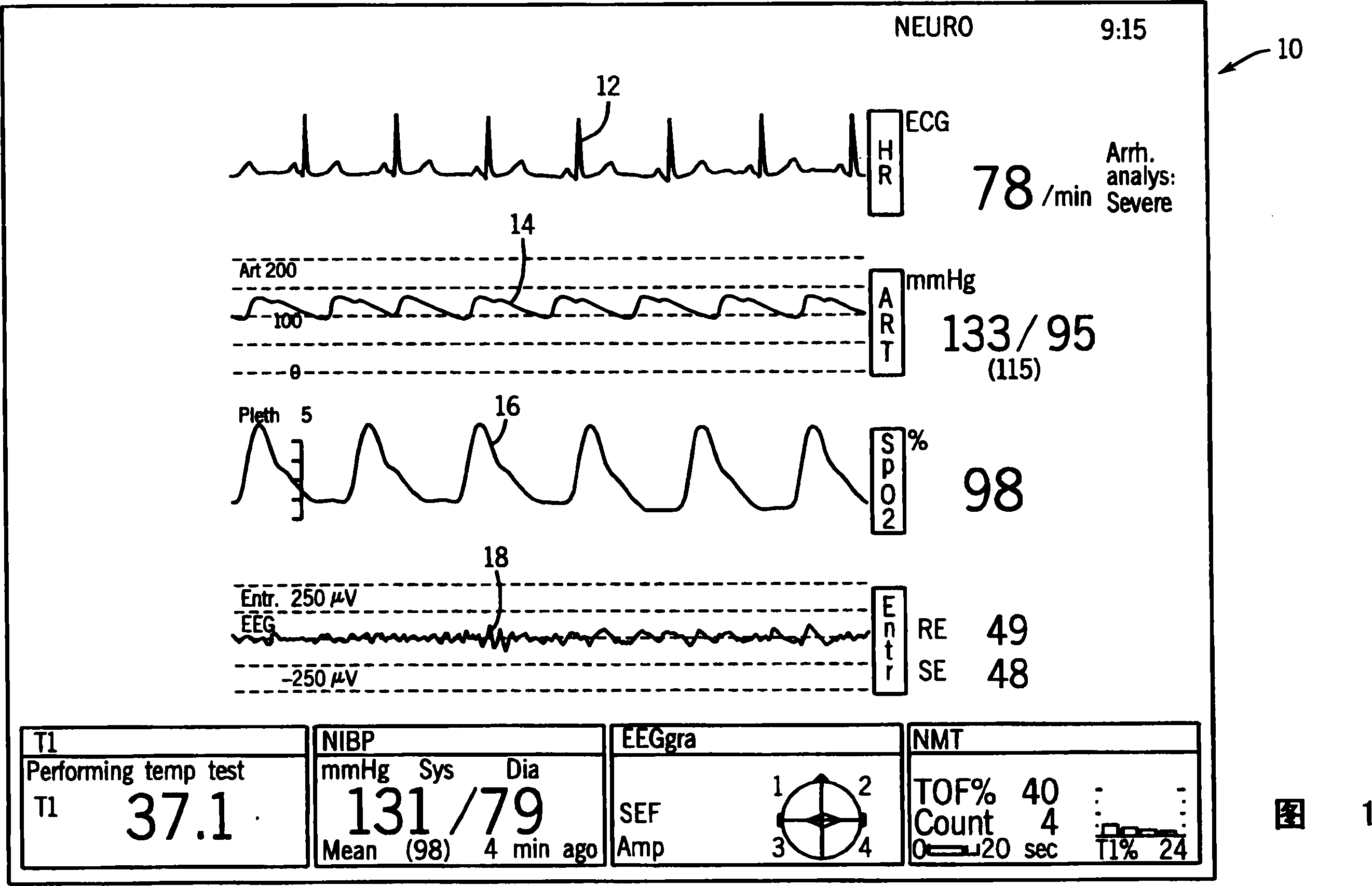
[0028] Referring first to FIG. 1 , there is shown a conventional display 10 as commonly seen in hemodynamic patient monitoring systems. The hemodynamic patient monitoring system has various uses and is widely used in operating rooms where anesthesia is used during surgery. As shown in FIG. 1 , a standard anesthesia display 10 graphically plots a set of hemodynamic measurements including at least one ECG curve 12 , invasive blood pressure curve 14 , oxygen saturation curve 16 , and entropy curve 18 . Each curve 12-16 is formed and presented in real-time on the display 10 and presents the user with information needed to monitor the patient during anesthesia. Each of the curves 12-18 is a conventional hemodynamic measurement and is widely used in the operating room environment.

[0029]Although the following description will make specific reference to a hemodynamic anesthesia monitoring system, it should be understood that the anesthesia monitoring system can be any type of pati...


PUM


Abstract

An apparatus and method for synchronizing the hemodynamic measurements (36,38,40) from a patient monitoring system (46) and the ultrasound images (32) from an ultrasound monitoring system (44). The synchronized images and measurements can be combined and displayed on a single display (48) that includes both the ultrasound images and the hemodynamic patient measurements. The combined display receives synchronized information (50) from both the ultrasound monitoring system and the hemodynamic patient monitoring system and displays the combined information on a single monitor. In addition to displaying the synchronized combined information, the system stores the synchronized information for later playback. The playback of the stored images and measurements is synchronized such that the clinician can be presented with the combined information in a usable manner.

Description

technical field [0001] The present invention relates to a cardiovascular monitoring system that synchronizes monitoring information from two cardiac monitoring devices and presents the information for viewing and analysis. More specifically, the present invention relates to a synchronization and storage system that combines information from an ultrasound monitoring system and a conventional hemodynamic patient monitoring unit such as is used during anesthesia procedures. Background technique [0002] Cross References to Related Applications [0003] This application is based upon and claims priority to US Provisional Patent Application Serial No. 60 / 610,473, filed September 16,2004. [0004] In modern operating rooms, anesthesiologists and surgeons utilize cardiovascular ultrasound units to monitor heart function during surgery. In cases where the anesthetist determines that the patient's cardiac status is weak or at risk, the clinician monitors the heart using ultrasound ...

Claims


Application Information

Patent Timeline
IPC IPC(8): A61B8/00A61B8/06
Inventor N·J·桑迪I·拉兹T·J·哈格尔洛姆
Owner GENERAL ELECTRIC CO
Why Eureka
Try Eureka