[0011]An object of the invention is to provide an improved method in comparison to the above, which, together with good planning, provides the best possible image quality of the magnetic resonance imaging dataset.
[0014]The invention can be applied with all types of magnetic resonance datasets, but preferably to at least three-dimensional magnetic resonance datasets. Examples include multi-slice imaging, with which a number of two-dimensional slices are acquired, three-dimensional static imaging, and three-dimensional dynamic imaging (with 3+1 (time) dimensions). Higher-dimensional magnetic resonance image data are also provided with other methods, for example multi-contrast-imaging. A series with two-dimensional images, the magnetic resonance data (raw data) of which are acquired interleaved with TSE sequences, provides significant sensitivity to motions.
[0023]In this case, it is obviously not necessary to discard all other motion status classes outside the selected motion status class. For example, an advantageous variant of the invention envisages that, in addition to the magnetic resonance data from the selected motion status class, the magnetic resonance data from at least one further motion status class satisfying a deviation criterion for the motion values with respect to the motion values of the data subsets of the selected motion status classes is also taken into account. This means it is also possible to use magnetic resonance data from motion status classes with a motion status or motion status range close enough to the motion status or motion status range of the selected motion status class. In this context, it is particularly advantageous for the magnetic resonance data of different motion status classes to be weighted, in particular with a weighting selected in dependence on a deviation of the motion values of its motion status class from those of the selected motion status class. Therefore, different motion statuses can be included in the reconstruction with different degrees of weighting, wherein the weighting is performed according to different criteria, but preferably based on the deviation of the motion status from the motion status of the selected motion status class. It is possible for different mathematical functions to be used in order to determine the weighting factors, for example in the sense of linear and / or exponential attenuation. A weighted consideration of further magnetic resonance data of this kind can, incidentally, be achieved particularly simply when iterative reconstruction of the magnetic resonance imaging dataset is performed. Overall, taking into consideration further magnetic resonance data from other motion status classes enables the signal-to-noise ratio to be improved since more magnetic resonance data is used for the reconstruction.
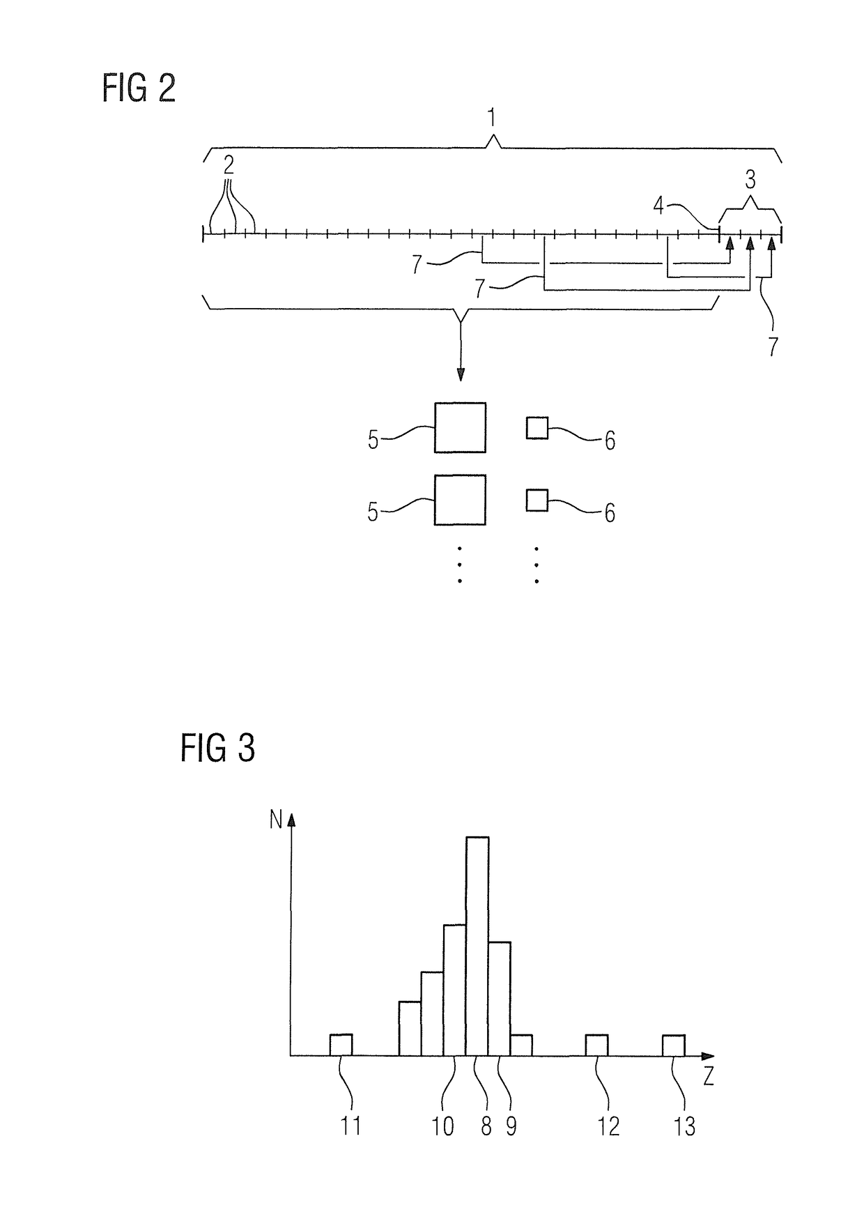
[0030]As explained above, it is expedient for the data subsets to be mutually supplementary with respect to the sampled data points in k-space so that, in all data subsets, or at least all data subsets to be acquired before a reserved sub-period, at least some different components of k-space assigned to the area to be examined are sampled. For example, at least two, in particular all, sub-periods for a predetermined region around the k-space center of k-space corresponding to the area to be examined, are sampled completely. This ensures, when the k-space center regions filled in all data subsets, magnetic resonance data from the k-space center are available for each motion status in question. It is also expedient if, taking together all and / or one predetermined component of the data subsets, a predetermined undersampling factor is obtained. If it is empirically known that, due to motions, usually 80% of the acquired magnetic resonance data can be used, it is expedient to arrange different sub-sampled components of k-space for the different sub-periods as equally distributed as possible. When using a component of magnetic resonance data of this kind, a specific, desired undersampling factor, for example the undersampling factor 2, is obtained. In this way, ultimately an acquisition concept is created that ensures uniform sampling of k-space and ensures a certain level of image quality.
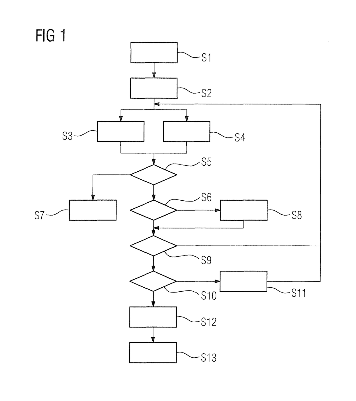
[0032]In a further, preferred embodiment of the invention, data subsets are selected as early as during the acquisition period and a preceding-image dataset is reconstructed on the basis thereof. When the preceding-image dataset satisfies a quality criterion describing a minimum image quality requirement, the acquisition process is aborted before the expiration of the acquisition period, and the most recently determined preceding-image dataset is used as a magnetic resonance imaging dataset. This embodiment is based on the concept that, during the arrangement and division of the acquisition period into sub-periods with respect to the magnetic resonance data to be acquired in the sub-periods, a certain buffer is to be created which, even upon the occurrence of magnetic resonance data that are non-correctable and / or cannot be taken into consideration with weighting, nevertheless enables the reconstruction of a sufficiently high-quality magnetic resonance imaging dataset. However, if it is the case that no motions occur over a lengthy period that would result in magnetic resonance data being discarded, and so a magnetic resonance imaging dataset of the desired quality can be reconstructed before the expiration of the acquisition period, it is no longer necessary also to acquire the remaining magnetic resonance data, and the acquisition can be aborted. Therefore, to this end, in real time, at least a portion of a preceding-image dataset is reconstructed from selected data subsets, or possibly further, data subsets that are weighted and / or taken into account subject to motion correction, and is checked with respect to the requisite image quality. If the image quality is sufficient, i.e. the quality criterion is satisfied, the acquisition can be aborted before the expiration of the acquisition period. In this case, it is particularly expedient for the evaluation of the quality criterion to be performed for a predetermined, two-dimensional slice in the area to be examined, since it is then easier to achieve real-time execution, thus enabling savings of calculation resources.
[0033]In another embodiment of the invention, during the acquisition process, if motion values are determined that satisfy an output criterion indicating excessive motion, that a predictive calculation shows will likely result in a failure to satisfy a quality target for the magnetic resonance imaging dataset, alerting information is emitted to notify the patient and / or an operator of the excessive motion. Therefore, if it is already evident from the motion values during the acquisition process that there is a risk that it will not be possible for the desired quality target to be achieved, the patient can be requested by the emission of the alerting information to reduce the number or extent of motions, if possible. In this way, it can be dynamically monitored during the acquisition process as to whether the patient is capable of sufficient cooperation for achieving an image of the necessary quality. Alerting information of this kind can also be provided to the operators, who can then calm the patient and encourage him or her to cooperate more fully. Hence, feedback of the motion monitoring is generated that can be used directly during the acquisition process.


