Method and arrangement relating to x-ray imaging

a x-ray and imaging method technology, applied in the field of x-ray imaging, can solve the problems of preventing the method from being used in cardiac imaging, breast imaging, etc., and achieve the effect of best possible image quality

Active Publication Date: 2007-05-24
PHILIPS DIGITAL MAMMOGRAPHY SWEDEN
View PDF4 Cites 53 Cited by
  • Summary
  • Abstract
  • Description
  • Claims
  • Application Information

AI Technical Summary

Benefits of technology

[0008] The object of the invention is to provide a novel X-ray apparatus for acquisition of dual energy images, using a single exposure, and best possible image quality for a given dose. The invention is based on prior art of multi-slit scanning using a photon counting line detector.

Problems solved by technology

High concentrations of a contrast agent make the tumor appear like a cloud in X-ray images, but unfortunately the cloud is super-imposed with anatomical structures, which may be strong enough to obscure the tumor.
In addition, the amount of injected contrast agents should be kept at moderate levels, since they may cause allergic reactions and kidney failure.
One problem of temporal subtraction is motion artifacts, which prevents the method from being used in cardiac imaging, and even breast imaging.

Method used


Image

  • Method and arrangement relating to x-ray imaging
  • Method and arrangement relating to x-ray imaging
  • Method and arrangement relating to x-ray imaging

Examples


Embodiment Construction

[0021] In the following description same reference signs refer to same parts throughout the drawings.

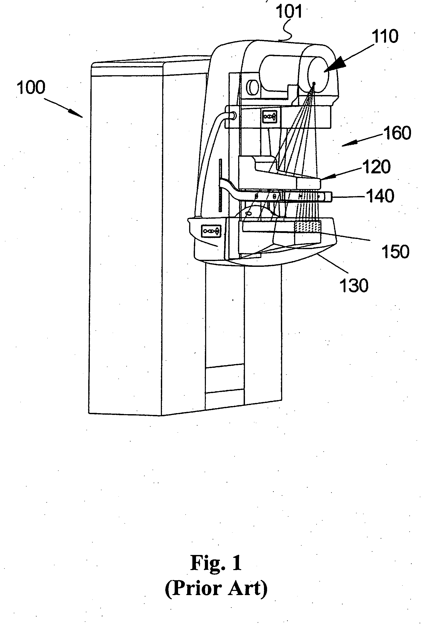
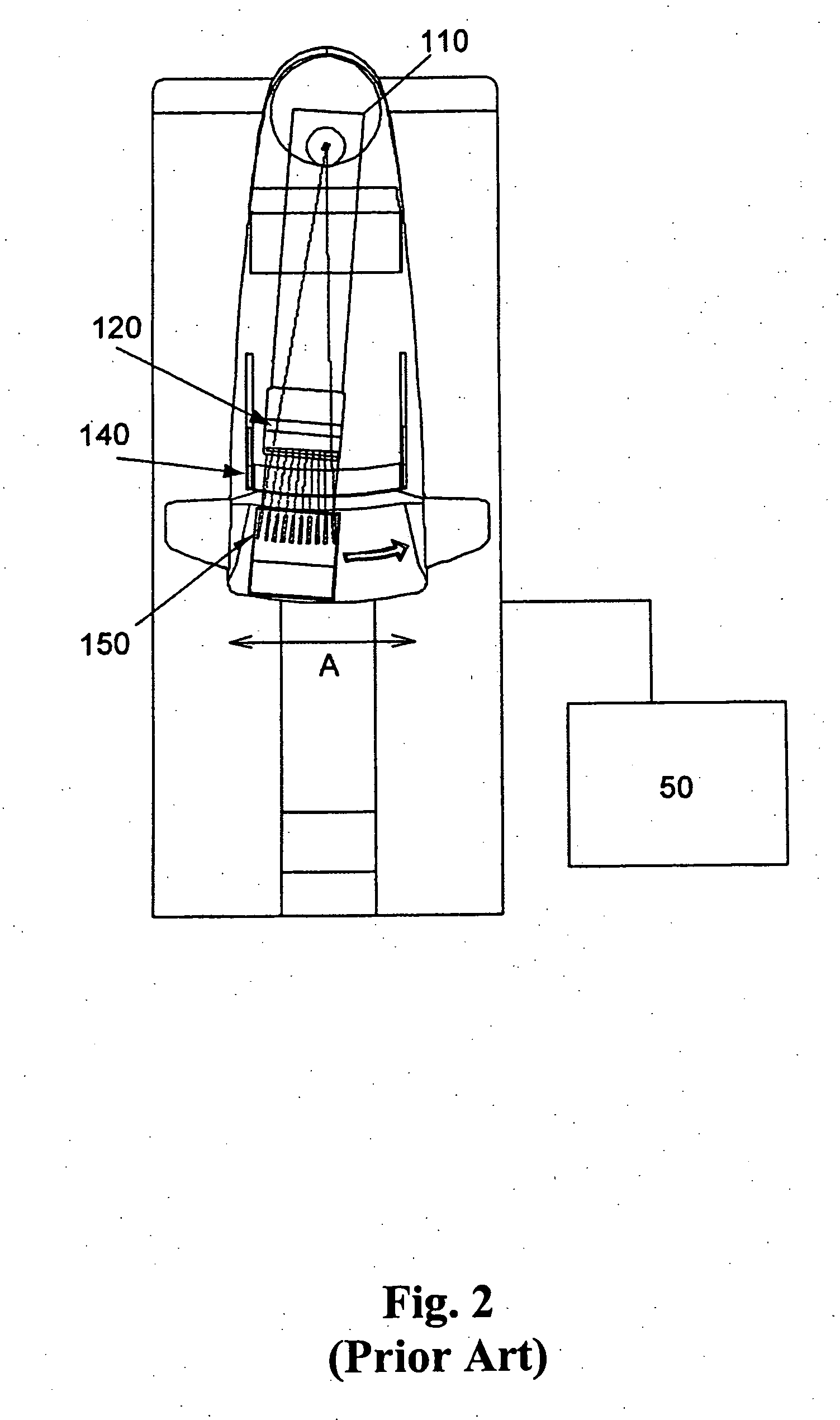
[0022] The preferred embodiment is based on arrangement and mechanical parts of prior art. The mechanical arrangement may be identical to prior art of multi-slit scanners, in FIGS. 1-2, but underlying system of electronics and software differ, according to the present invention.

[0023] A pulse counter is defined to be any scalar register, which is setup to increment or decrement its value for each received pulse. The step for increment or decrement may vary. The register representation may be integer, floating point or similar. In the most preferred embodiment, the counter is an integer value, which is incremented by a fixed step for each pulse, and never decremented, and the step is equal to one.

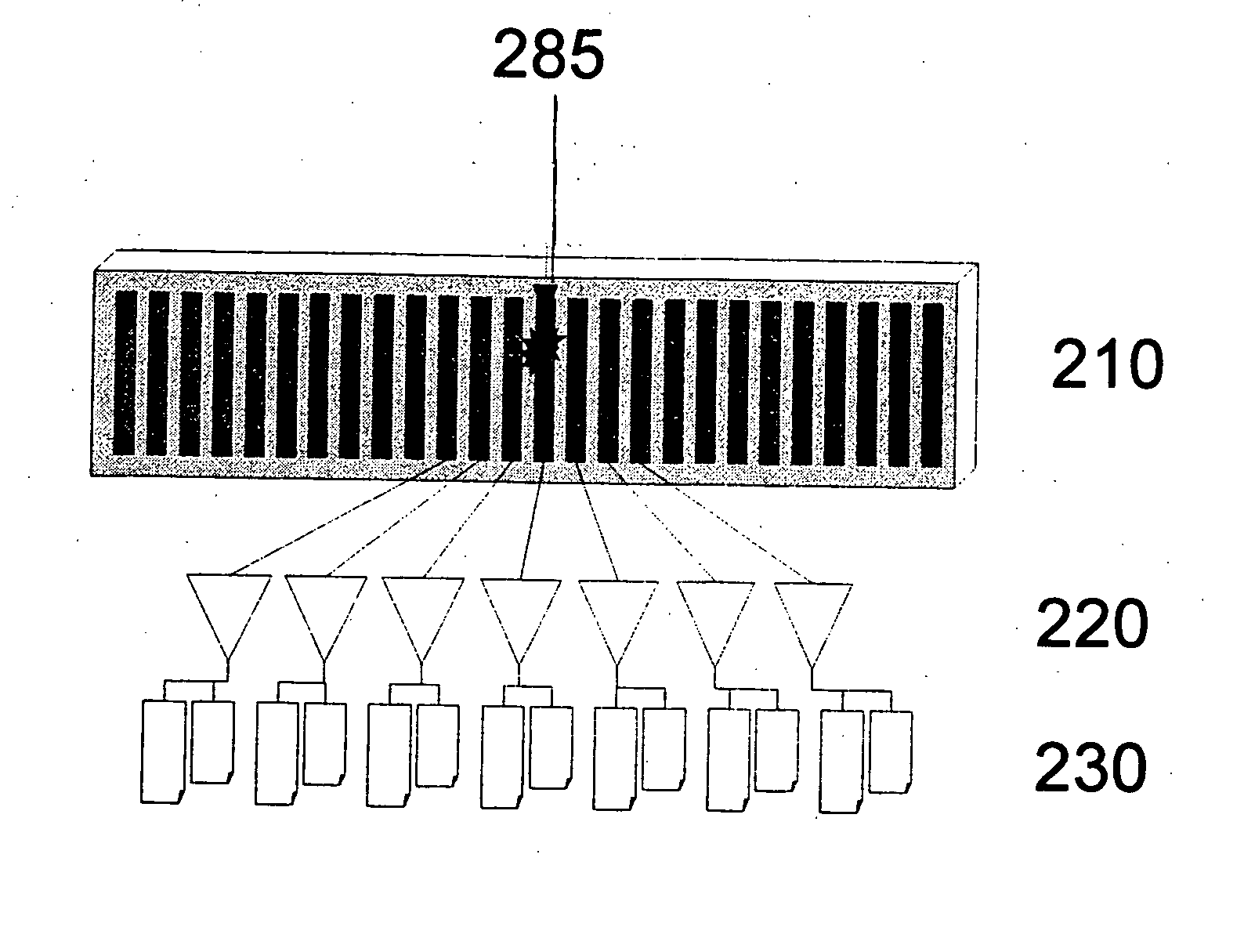
[0024]FIG. 4 illustrates a line detector according to one embodiment of the invention. The line detector comprises a silicon detector 210, which is an array of photon conversion channel el...


PUM


Abstract

An X-ray apparatus is provided for acquisition of images containing spectral information. An X-ray source and a collimator having multiple slits are operable together with a set of line detectors, the line detectors including linear arrays of photon counting channels. Each of the channels includes a photon conversion channel element which is operable to convert photons to electric pulses. A plurality of pulse counters are operable to count pulses in a plurality of different ranges of pulse strength, where the strength of a pulse depends on the energy of the photon. Further, an arrangement is included for an energy subtracting operation.

Description

TECHNICAL FIELD OF THE INVENTION [0001] The present invention relates to a method and arrangement in X-ray imaging, in particular dual energy, contrast agents, photon counting detectors, and multi-slit scanners for mammography. BACKGROUND OF THE INVENTION [0002] The tissue around a typical cancer tumor is characterized by angiogenesis, which is a growth of nearby blood vessels to supply the growing tumor. Most likely, the tumor contains a lot more blood than healthy tissue. A widely used method of detection is to inject contrast agents and acquire images, either using x-ray imaging or magnetic resonance imaging (MRI). For X-ray imaging, the most widely used contrast agents are based on iodine, which attenuate x-rays more than normal tissue. High concentrations of a contrast agent make the tumor appear like a cloud in X-ray images, but unfortunately the cloud is super-imposed with anatomical structures, which may be strong enough to obscure the tumor. In addition, the amount of injec...

Claims


Application Information

Patent Timeline
Patent Type & Authority Applications(United States)
IPC IPC(8): G01T1/24
CPCA61B6/4241A61B6/482A61B6/583G01T1/2928A61B6/502
Inventor DANIELSSON, MATSLUNDQVIST, MATS
Owner PHILIPS DIGITAL MAMMOGRAPHY SWEDEN
Why Eureka
Try Eureka