Method for performing stereotactic brain surgery using 3D geometric modeling

a geometric modeling and stereotactic brain technology, applied in the field of stereotactic brain surgery using 3d geometric modeling, can solve the problems of significant errors and distortions in the subject's measurements, lack of precise definition of the neutral position of the 3d orientation of the human head, and no existing method yields sufficient accuracy in defining the neutral, etc., to achieve accurate, reliable, user-friendly and portable

Inactive Publication Date: 2018-10-02
ALBERT DAVYDOV
View PDF8 Cites 0 Cited by
  • Summary
  • Abstract
  • Description
  • Claims
  • Application Information

AI Technical Summary

Benefits of technology

[0006]It is an object of the present invention to provide a method of performing head motion and orientation measurements that are accurate, reliable, user-friendly and portable with low-cost.

Problems solved by technology

However, none of the existing methods yield a sufficient accuracy in defining the neutral position of a human head in three-dimensions.
This lack of a precise definition of the neutral position of the 3D orientation of the human head is a serious disadvantage in current 3D geometric modeling, especially when one needs to determine some specific measurements and orientation of the human head during motion.
In certain applications, for example in dental arts and in stereotactic brain surgery, the existing methods result in significant errors and distortions in the subject's measurements and, consequently, in the resulting 3D model.
Because the surgeon relies on this image during the surgery, this imprecision sometimes results in severe damage to the patient's brain.

Method used


Image

  • Method for performing stereotactic brain surgery using 3D geometric modeling
  • Method for performing stereotactic brain surgery using 3D geometric modeling
  • Method for performing stereotactic brain surgery using 3D geometric modeling

Examples


Embodiment Construction

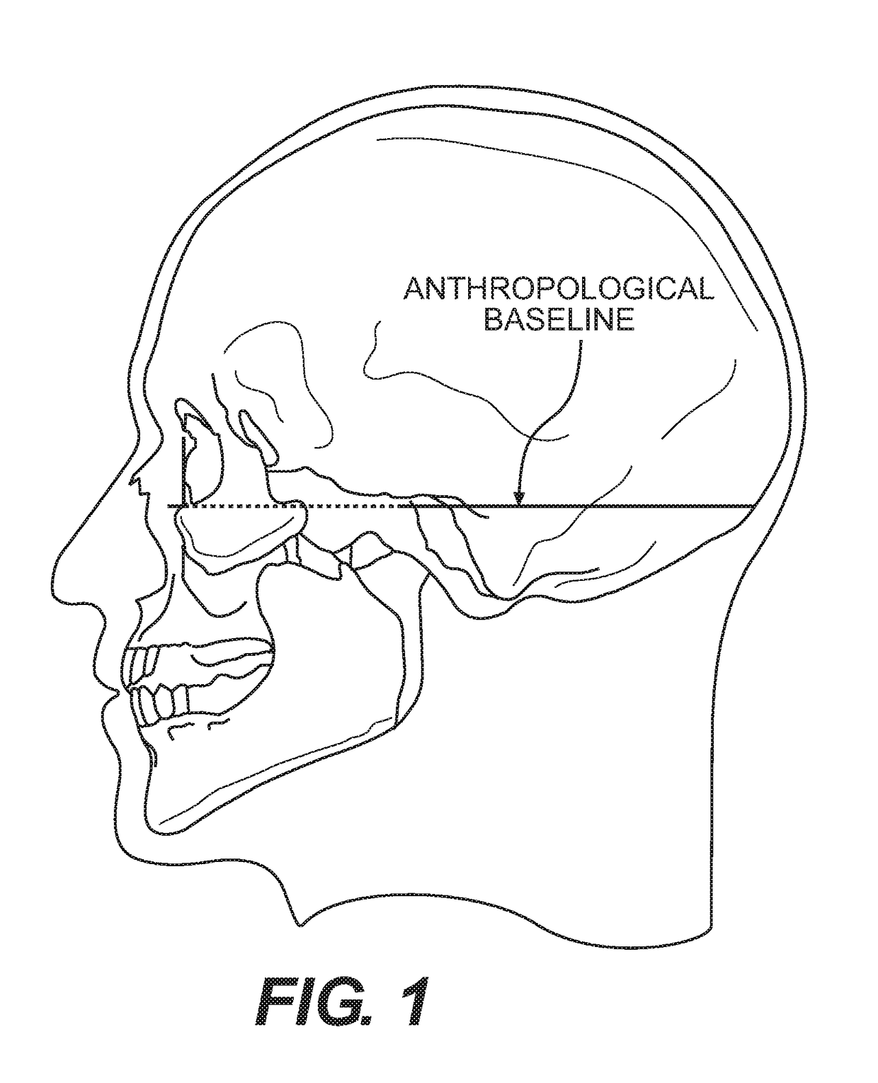
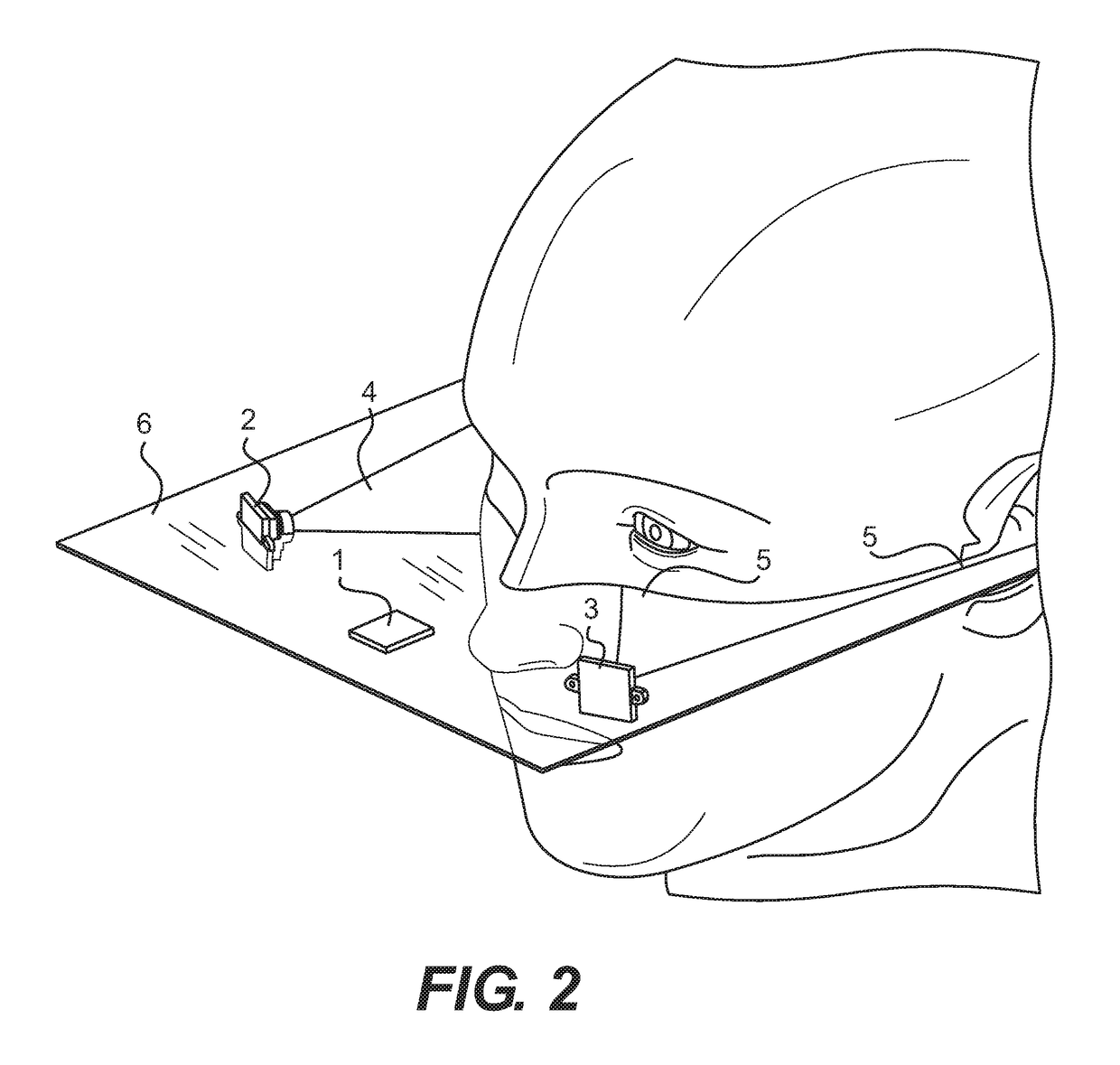
[0018]As illustrated the attached Figures, the method and apparatus of the present invention are used to determine a correct neutral position of the 3d orientation of a human head. Once such correct neutral position is determined, it can be used for an unambiguous definition of the 3D orientation of the human head and for head orientation during motion.

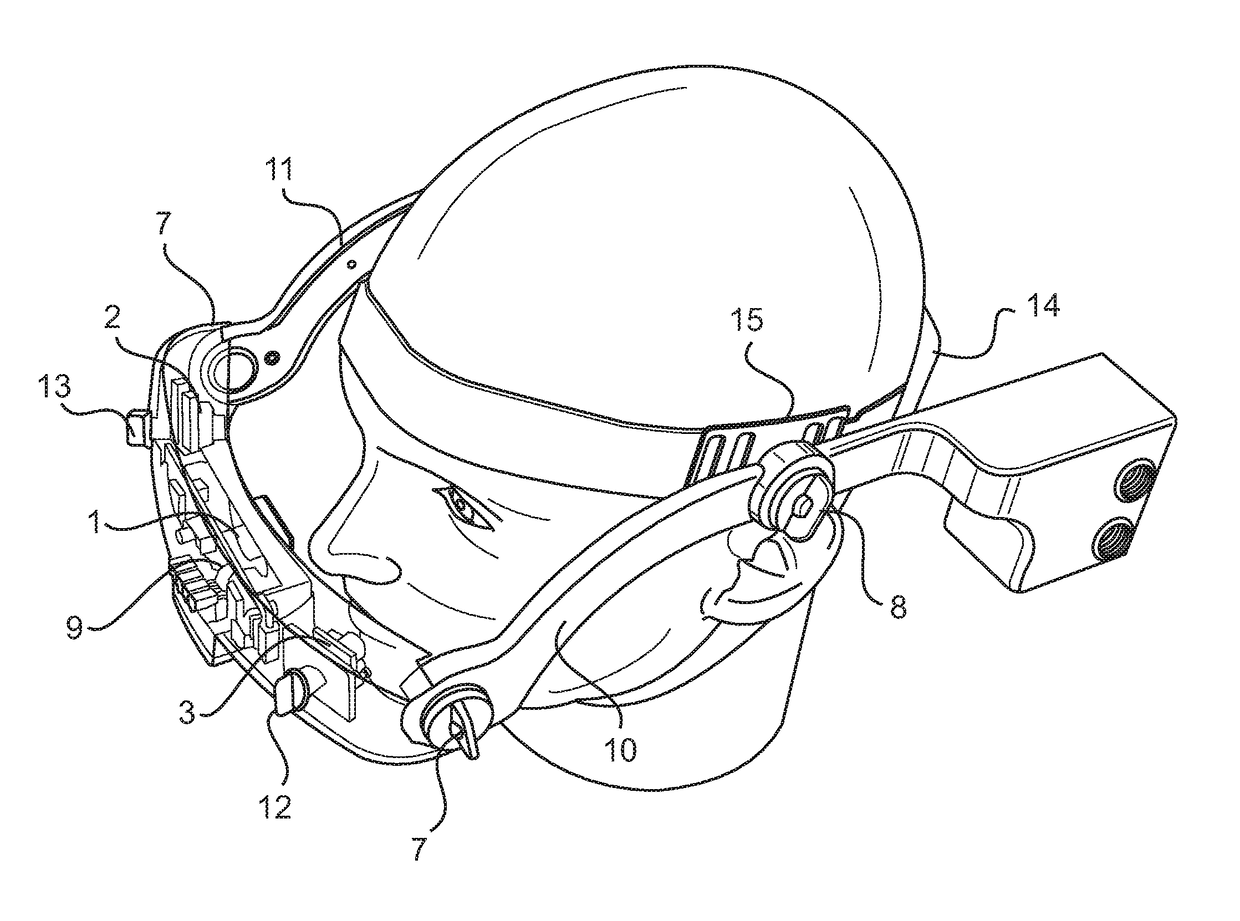
[0019]Elements of the apparatus for determining the correct neutral position of the 3d orientation of human head are shown in FIGS. 2-3. In accordance with the preferred embodiment, the headgear unit 100 has a plastic frame 16 with two temple elements 10, 11 and a front housing 18. At least one 3D compass 1 with its electronic hardware 9 are secured within the front housing 18. The 3D compass 1 is an integrated compass module for heading computation and calibration of magnetic distortions. The module combines 3-axis magneto-resistive sensors and 3-axis micro electro-mechanical systems accelerometers, analog and digital support circuit...


PUM


Abstract

A method for performing a stereotactic brain surgery. The method includes first interposing a left and a right anthropological baselines on a patient's face, each trago-orbital anthropological baseline connecting an orbitale point and an auricular point on a corresponding side of the patient's face. A headgear unit having a pair of temple elements and a front housing, where the front housing includes at least two cameras and a 3D compass, is then positioned on a patient's head. Each of the two cameras is adjusted such that its view field picks up one of the two trago-orbital anthropological baselines on the patient's face. These view fields of the two cameras are then used to construct a Human Skull Base Plane as a plane formed by the left and right anthropological baselines when these anthropological baselines are parallel and aligned. The same view fields of the two cameras are also used to identify a precise center of the patient's face, and a center of the front housing is then positioned in alignment with the center of the patient's face. The 3D compass is then positioned into a measuring plane coinciding with the Human Skull Base Plane, and is used to quantitatively measure a three-dimensional orientation of the patient's head at the time when the 3D compass is positioned within the measuring plane coinciding with the constructed Human Skull Base Plane. An image of a patient's brain is then obtained and stored in correlation with the measured three-dimensional orientation of the patient's head. Prior to the surgery, the patient's head is positioned into the same position in which the image was taken. A stereotactic brain surgery is then performed using the stored image.

Description

BACKGROUND OF THE INVENTION[0001]This application and its disclosure generally relate to the field of determining orientation of a human head in three-dimensional geometric modeling and then using this orientation in stereotactic brain surgery.[0002]Head orientation measurements in 3D geometric modeling are currently based on a combination of measurements determined by different gyroscopes, accelerometers and electronic compasses. For example, one such system is based on measurements provided by a three-axis accelerometer and a resonator gyroscope mounted on a headgear.[0003]Other methods and measuring systems, currently in use, utilize dual inclinometers, bubble goniometers, radiographs, compass technology, visual estimation, ultrasound, geometric methods, digital optoelectronic instruments, computerized kinematic analysis using passive markers and infrared TV cameras, MRI or sensors attached to the subject's head for head orientation determination. However, none of the existing me...

Claims


Application Information

Patent Timeline
Patent Type & Authority Patents(United States)
IPC IPC(8): A61B90/00H04N13/243G06T7/73A61B90/14A61B5/11A61B5/107A61B5/00A61B90/50
CPCG06T7/74H04N13/243A61B90/14A61B5/1077A61B5/1079A61B5/1122A61B5/1128A61B5/6803A61B90/361A61B90/37A61B5/0077G06T2207/30196A61B2090/371A61B2090/502G06T2207/10028G06T2207/30016
Inventor DAVYDOV, ALBERTUSOV, PETER
Owner ALBERT DAVYDOV
Why Eureka
Try Eureka