Analysis methods of ultrasound echo signals based on statistics of scatterer distributions

an analysis method and ultrasound echo signal technology, applied in the field of system and method for analyzing ultrasound echo signal, can solve the problems of inability to characterize pathological characteristics with low contrast, weak echo signal received by ultrasound probe, and inability to capture images captured by different ultrasound systems

Active Publication Date: 2018-10-23
AMCAD BIOMED CORP
View PDF5 Cites 0 Cited by
  • Summary
  • Abstract
  • Description
  • Claims
  • Application Information

AI Technical Summary

Benefits of technology

[0013]The system is based on the above method and performs statistical analysis of signal image values of ultrasound image data through multiple statistics of scatterer distributions, so as to evaluate distribution and arrangement of scatterers within a tissue, thereby assisting in providing clinical information on tissue state. The system comprises a capturing device, an analyzing unit, and a display unit.

Problems solved by technology

Because echo signals received by an ultrasound probe is very weak and complex, it is necessary to display an image through many signal processing steps.
However, images captured by different ultrasound systems may have different quality in the same lesions.
The reason is that grayscale ultrasound data exhibits a granular pattern of white and dark spots, named speckle, so some pathological characteristics with low contrast cannot be characterized.
The speckles dim an image of a tiny structure and reduce the contrast and resolution of the ultrasound image.
Although this is beneficial for clinical anatomical observation, another problem is formed because that physiological or pathological information may be lost.
That is why a traditional grayscale image cannot provide the information of characteristics of the tissue.

Method used


Image

  • Analysis methods of ultrasound echo signals based on statistics of scatterer distributions
  • Analysis methods of ultrasound echo signals based on statistics of scatterer distributions
  • Analysis methods of ultrasound echo signals based on statistics of scatterer distributions

Examples


Embodiment Construction

[0030]In order to fully understand the effects of the present invention, preferred embodiments are described below in combination with the accompanying drawings.

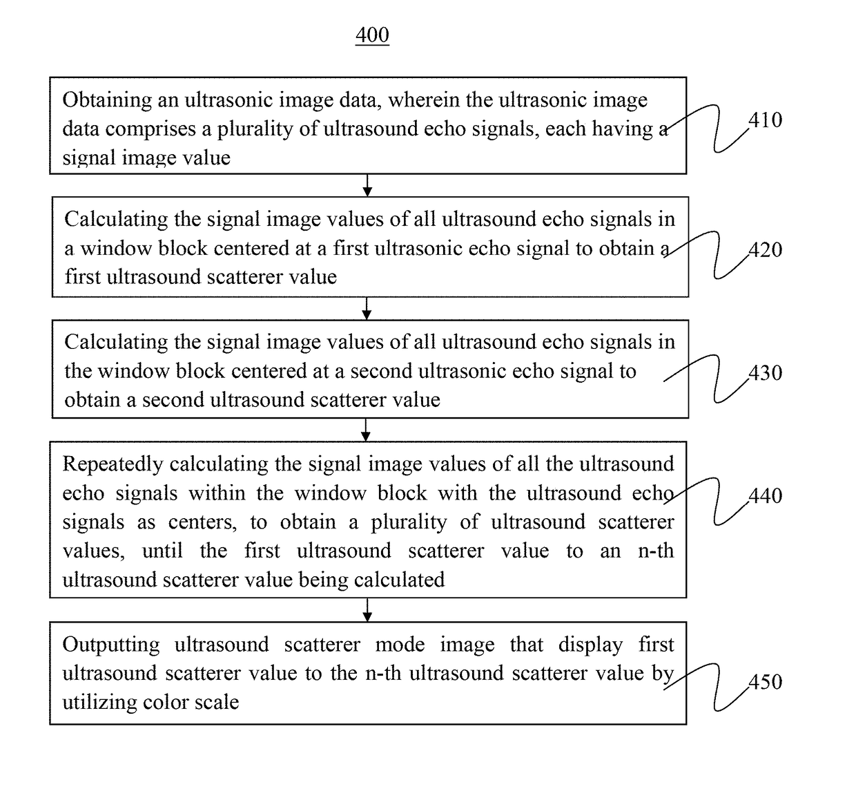
[0031]The present invention provides a method and a system for analyzing ultrasound echo signals based on statistics of scatterer distributions, wherein analysis of signal image values of ultrasound image data are performed based on statistics of scatterer distributions, so as to evaluate distribution and arrangement of scatterers within a tissue, thereby assisting in tissue characterization and providing clinical information on tissue state.

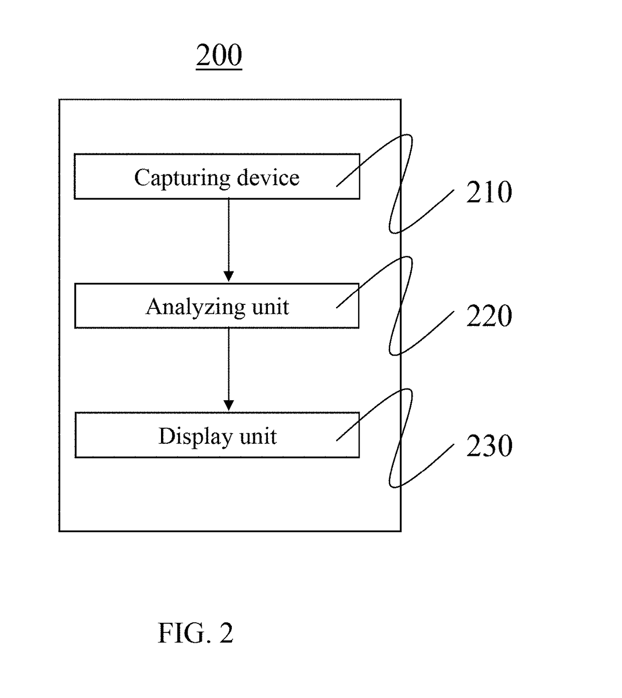
[0032]A schematic diagram of a system designed for analyzing ultrasound echo signals based on statistics of scatterer distributions is shown in FIG. 2. An ultrasound image system 200 of the present invention comprises a capturing device 210, an analyzing unit 220, and a display unit 230. A schematic view for illustrating operation of a moving window block 330 in an ultrasound image accor...


PUM


Abstract

The invention provides an analysis method of ultrasound echo signals based on statistics of scatterer distributions. The beginning of steps, choosing an ultrasound echo signal as a center, and calculating the signal image values of all ultrasound echo signals within a window block in an ultrasound image data to obtain an ultrasound scatterer value. Then, choosing another ultrasound echo signal as the center to repeat the previous steps until all of ultrasound echo signals may be calculated. The interval between each of ultrasound echo signal is one point distance. Finally, to output an ultrasound scatterer mode image with all ultrasound scatterer values by utilizing color scale. The ultrasound scatterer mode image can assist doctor to confirm the relative region of lesion in a target organ.

Description

TECHNICAL FIELD[0001]The present invention relates to a system and a method for analyzing ultrasound echo signals, and in particular, to a system and a method for analyzing ultrasound echo signals based on statistics of scatterer distributions.BACKGROUND ART[0002]A flow chart 100 of generating an ultrasound grayscale image according to the prior art is shown FIG. 1. Because echo signals received by an ultrasound probe is very weak and complex, it is necessary to display an image through many signal processing steps. In step 110, echo signals data are acquired from an ultrasound probe. However, echo signals still vibrate up and down. In step 120, called a signal demodulation step, negative-phase signals are converted into positive-phase signals, and waveform shape are taken for generating ultrasound envelope data. Because the energy of echo signals is attenuated with the depth of a tissue, the echo signal is required to compensate and amplify through depth gain compensation. Also, be...

Claims


Application Information

Patent Timeline
Patent Type & Authority Patents(United States)
IPC IPC(8): A61B8/08A61B8/00A61B8/14
CPCA61B8/5215A61B8/461A61B8/14A61B8/0891A61B8/5207A61B8/5269G01S15/8977G01S7/52077
Inventor CHEN, ARGONWANG, YU-HSINHUANG, KUO-CHENCHEN, JIA-JIUN
Owner AMCAD BIOMED CORP
Why Eureka
Try Eureka