Integrated membrane sensor for rapid molecular detection

a sensor and integrated technology, applied in the field of microfluidic membrane sensing technology, can solve the problems of limiting the usefulness of field applications, lack of actionable information, and inability to be used as early diagnostic markers, etc., to improve sensitivity, reduce assay cost, and improve sensitivity. the effect of time and stability

Active Publication Date: 2019-04-02
UNIV OF NOTRE DAME DU LAC
View PDF8 Cites 0 Cited by
  • Summary
  • Abstract
  • Description
  • Claims
  • Application Information

AI Technical Summary

Benefits of technology

[0015]In some embodiments disclosed herein is a microfluidic sensing device and method wherein the device is a nucleic acid sensing platform capable of rapid, high throughput, accurate, and reliable identification of pathogen-specific nucleic acids in a field-expedient format. Molecular detection is accomplished without amplification of nucleic acid, using an extremely sensitive nanomembrane-based electrokinetic sensor, capable of detecting DNA or RNA target sequences. Without the need for thermocycling or a dedicated heat block, the detection platform described herein is rapid, sensitive, low-cost, easy to use, and portable, making it amenable to point-of-care applications. The device described herein is therefore capable of evaluating human clinical specimens for the presence of pathogen-specific RNA and thus provide critical information during a rapid response to an emerging infectious disease outbreak or a bioterrorism event. Development of a cost-effective, point-of-care diagnostic instrument for rapid, sensitive and specific detection of DENV from patients can help to circumvent many inherent difficulties related to control of dengue fever and severe dengue.
[0017]DENV is an RNA virus with four different serotypes (DENV-1, 2, 3, 4). While the RNA of the virus is stable within the viral shell, the chances of RNA degradation increase during the extraction phase if exposed to ubiquitous RNase enzymes. RNA detection therefore represents a higher bar than DNA, which the platform can overcome. DENV is also a National Institute of Allergy and Infectious Disease (NIAID) Category A pathogen, and a rapidly emerging global public health threat. Early diagnosis of dengue therefore has significant clinical importance, and can help facilitate appropriate supportive therapies to reduce DENV associated morbidity and mortality. In austere environments where RNA preservation cannot be easily carried out, an integrated diagnostic assay capable of RNA extraction and detection with minimum human intervention represents an urgent global health need.
[0020]The pre-treatment unit for a real or raw sample (for example, blood, saliva, water, or urine) lyses the cells, bacteria, viruses, and other bioparticles to release the biomarkers. Upon application of a DC field, the negatively charged biomolecules are pulled out of the lysate through a thin layer of agarose gel towards the positively biased electrode. The agarose gel serves as a mechanical barrier between the sample chamber and the main channel on the chip. Nanoporous heterogeneous cation exchange membranes contain negative fixed charge that does not allow the passage of negatively charged molecules (nucleic acids / proteins) due to both steric effect and strong electrostatic repulsion. The pretreatment unit thus allows selective separation of negatively charged biomolecules from the lysate and their containment in another fluidic chamber for further downstream processing.
[0023]Different designs of the three aforementioned units and the fluidic structures are first fabricated as stand-alone units and then put together using adhesives. The fluidic structures are made in plastic sheets that are assembled by thermal fusing. The three units are made through a combination of molding processes for precise membrane positioning and various auxiliary techniques (micromachining, drilling, etc.) for the whole unit assembly. All the techniques are inexpensive and easy to use without requiring highly trained personnel. The platform is novel in that it not only extracts, concentrates and detects on-chip, but is also capable and amenable to change / switch out all the three components including a variety of potential biosensors.
[0025]The integrated nucleic acid detection platform circumvents the issues associated with electrochemical / fluorescent-based sensing techniques by reducing assay cost, assay time, improving sensitivity, stability and portability for point-of-care applications. As described herein, and as will be readily apparent from the following descriptions of the drawing and exemplary embodiments, the platform enables molecular detection in a miniature format, amenable to use in any setting where such detection is required—be it near the patients' bedside at a sophisticated hospital, or in a clinic in a low resource setting, or by first responders investigating a biological event or a medic at a military base.

Problems solved by technology

However, these traditional methods of viral detection have a number of disadvantages that limit their usefulness for field applications.
Results from virus isolation may take days and thus offer little actionable information during the course of an infection.
ELISA techniques, although not time-consuming, typically requires 4-7 days for the body to produce appreciable antibodies by immunological response after virus infection and thus cannot be used as an early diagnostic marker.
Although the FDA recently cleared the first molecular test (CDC DENV-1-4 RT-PCR Assay) for dengue detection, the Applied Biosystems 7500 Fast Dx Real-time PCR Instrument is not portable and thus precious time is spent transporting specimens (2-3 hours of test time+potentially 24-48 hours of sample delivery time) and waiting for the results to return.
A 2-3 hour result is a significant improvement but the complexity of such systems including its lack of portability, expensive reagents and requirement of technical personnel for operation become an economic disadvantage.
Some optical detection techniques, for instance surface plasmon resonance imaging (SPRi) microarray (Nelson 2002) and mass-sensitive detection such as microcantilever designs (Zhang 2006) are label free, but require expensive equipment and have signal instability issues.
Although these optical detection methods provide reasonable detection limits down to an impressive single molecule level, they all require costly, bulky optical systems.
Several label-free molecular sensing technologies like electrochemical sensors that amplify signal using redox reporters enhance detection sensitivity, yet are hampered by instability of the electrochemical signal and difficulty in calibration (Bakker and Qin 2006, Umezawa and Aoki 2004).
They are also expensive since they require fabrication of microelectrodes.
Most importantly, the largest drawback of all electrode sensors is their long assay time due to diffusion-based transport of large target molecules like DNA / RNA to the electrode surface (Nkodo 2001).
However, this electrophoretic concentration technique is highly non-specific and the elevated voltage can produce undesirable Faradaic reactions for high ionic strength buffers resulting in false current or voltage signals.
Therefore, the major disadvantages of existing detection technologies are 1) time consuming, 2) expensive, 3) require trained personnel and 4) not suitable for field diagnostics.
Thus, the lack of availability of a point-of-care diagnostic platform has made the detection of viruses in human sera particularly cumbersome in endemic areas.

Method used


Image

  • Integrated membrane sensor for rapid molecular detection
  • Integrated membrane sensor for rapid molecular detection
  • Integrated membrane sensor for rapid molecular detection

Examples


examplei

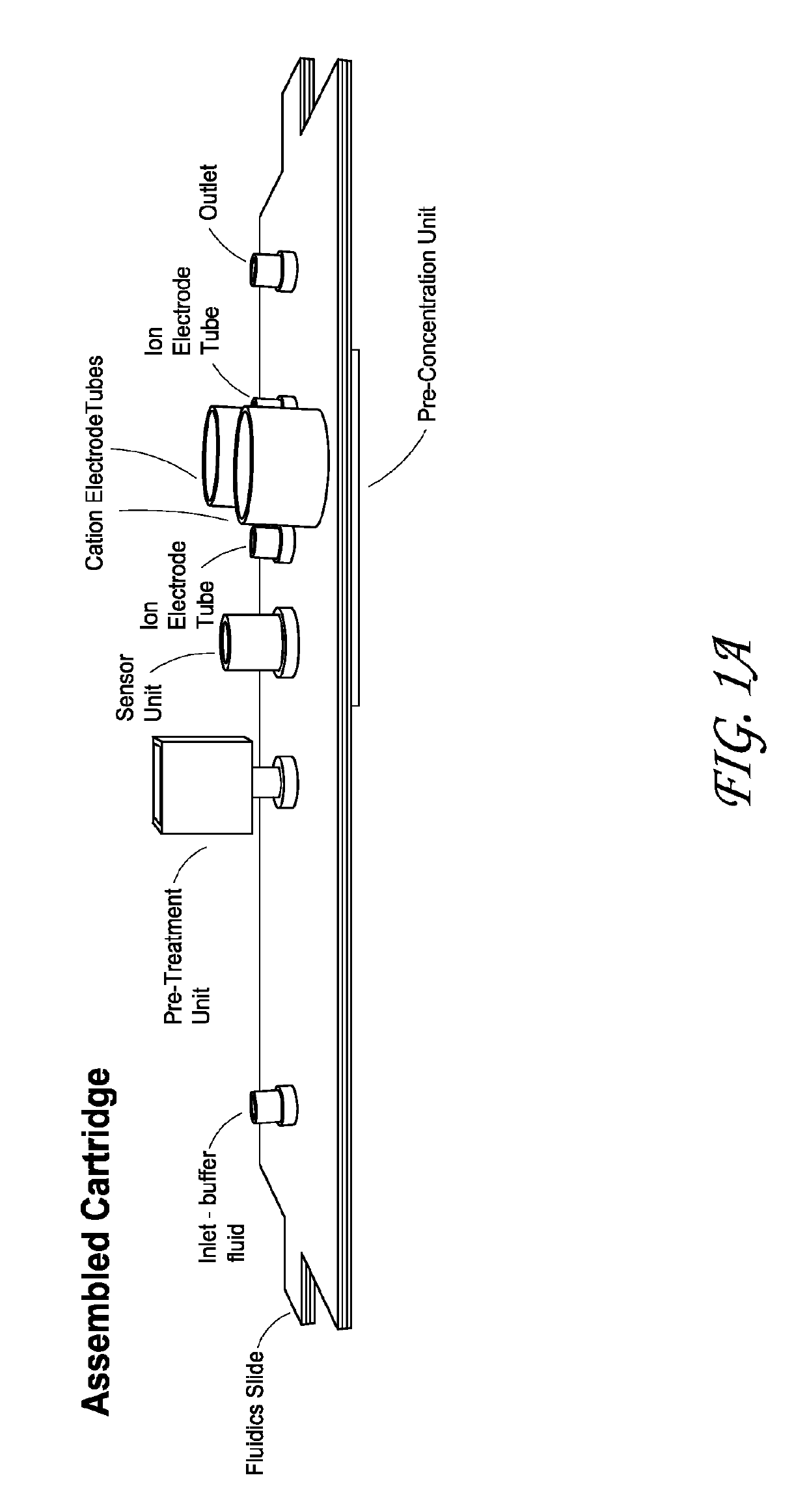
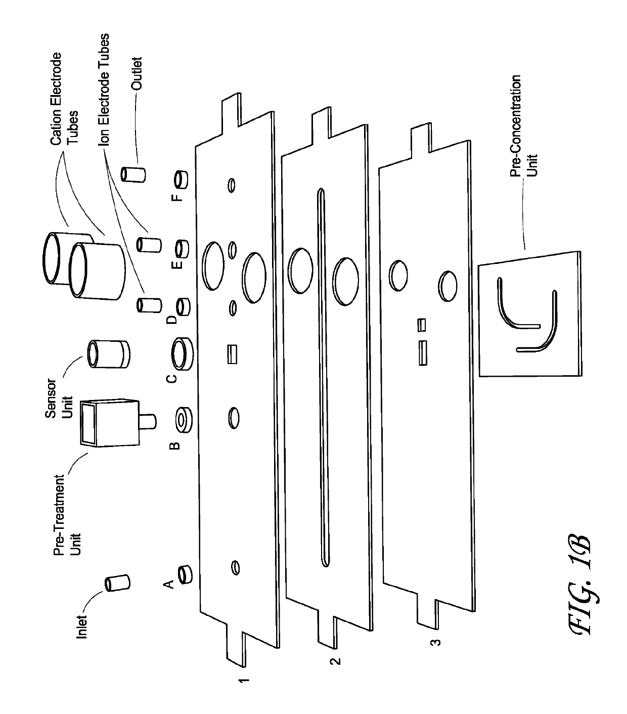
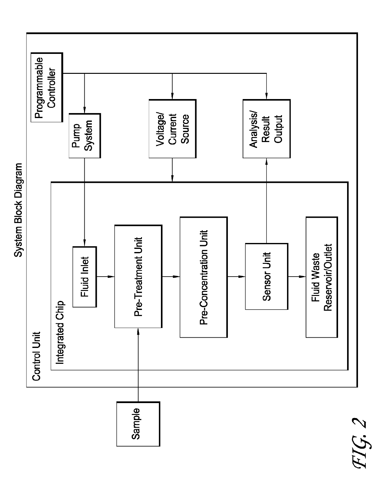
ure of an Integrated Chip

[0155]In some embodiments of the integrated chip, the integrated chip comprises a reference electrode, a source electrode, a reservoir, an anion membrane, a resin, a photoreactive benzophenone-3,30,4,40-tetracarboxylic acid (powder)—1 mg (measured on scale) in 10 μL of DI water maintaining a pH of 6-7, sodium hydroxide—solid pellet (Fisher Scientific 500 gm). The integrated chip is fabricated comprising a sensor disc, wherein once the sensor disc is out of the mold, measure I-V curve (same step up as when experiment is run, must be identical), a reservoir, an assembled sensor disc & reservoir.

exampleii

lization of the Membrane

[0156]The membrane may be functionalized as described previously. A functionalization of the membrane may be performed prior to sensor disc step or after assembly of disc and reservoir, however, it is better to do so after because there are fewer chances of contamination afterward. The membrane may be carboxylated by: 1) Dissolving 1 mg powder photoreactive benzophenone 3,3,4,4 in 10 μL of DI water; 2) Adding dissolved sodium hydroxide to achieve a pH of 6-7 (roughly 2 μL); 3) Placing a drop onto the membrane, let sit for 5 minutes; 4) Exposing to UV for 10 minutes, atmosphere in UV box in nitrogen (efficiency of reaction will increase with the gas); 5) Put in DI water and shake it to wash membrane, can use equipment to assist with this like a vortex, vibrating, or rotating machine; 6) Repeat last three steps two more times; 7) Leave in 0.1×PBS overnight. This swells the membrane; 8) Measure I-V curve (same step up as when experiment is run, must be identical...

exampleiii

of the Target Probe

[0158]Attachment of the probe to the functionalized membrane may be achieved as described herein. Following the prior I-V curve measurement, soak sensor unit in pH 3 for one hour. Following the one hour incubation: 1) Wash with 0.1×PBS; 2) Wash membrane surface with MES buffer (mixed at pH 5.5) three times, can use vortex machine, rotator, etc. for assistance; 3) Add 0.19 mg of or final mixture amount of 0.4 molar, powder (refrigerated, did not get name of chemical b / c camera did not focus); 4) Other chemistry will work to allow bonding of probe; 5) Leave drop for 30 minutes on top of ‘dot’ membrane area; 6) Leave inside some moist box / chamber so sensor unit does not dry out during the 30 minutes; 7) Wash with MES buffer; 8) Apply DNA probe with NH21 group (needs clarification), DNA probe modified with amine group; 9) Generally use 1 micromolar concentration of that DNA group in 0.1×PBS; 10) Put in the moisture chamber and leave overnight; 11) Wash with 0.1×PBS; a...


PUM

PropertyMeasurementUnit
timeaaaaaaaaaa
timeaaaaaaaaaa
areaaaaaaaaaaa

Abstract

Disclosed are methods, compositions, and devices for an integrated, heterogenerous ion-exchange membrane-based plastic microfluidic biochip platform that can be used to detect multiple diagnostic markers present in real samples. Its various components can be easily integrated in a modular fashion for different applications. Automated control allows sequential and dynamic activation of different components on the chip. The integrated platform consists of three units and is designed to execute the following functions: (i) separation of the target biomolecules from the real sample, (ii) localizing and concentrating the targeted molecules at a specific location in the microfluidic chip, and (iii) detection of the targeted molecules using hybridization / docking events against a complementary ssDNA oligoprobe sequence or a specific antibody.

Description

CROSS-REFERENCE TO RELATED APPLICATIONS[0001]This application is a U.S. National Phase Application of PCT International Application Number PCT / US2015 / 032079, filed on May 21, 2015, designating the United States of America and published in the English language, which is an International Application of and claims the benefit of U.S. Provisional Application No. 62 / 002,098, filed May 22, 2014, U.S. Provisional Application No. 62 / 051,606, filed Sep. 17, 2104, and U.S. Provisional Application No. 62 / 075,647, filed Nov. 5, 2014, the disclosures of which are hereby expressly incorporated by reference in their entireties.BACKGROUND OF THE INVENTION[0002]Field of the Invention[0003]The disclosure relates to the field of microfluidic membrane sensing technology and more particularly, to methods and apparatus for nanomembrane based nucleic acid sensing platform.[0004]Description of the Related Art[0005]Dengue virus (DENV) is the world's fastest spreading tropical disease with an estimated 390 m...

Claims


Application Information

Patent Timeline
Patent Type & Authority Patents(United States)
IPC IPC(8): B01L3/00G01N27/327G01N27/333G01N33/487G01N27/447G01N1/40
CPCG01N33/48721B01L3/502753G01N27/3276G01N27/44743G01N27/44791G01N2001/4011B01L2300/0681B01L2400/0415B01L2400/0418B01L2400/0421B01L2400/0487B01L2300/0645
Inventor SLOUKA, ZDENEKSENAPATI, SATYAJYOTISHAH, SUNNY S.CHANG, HSUEH-CHIA
Owner UNIV OF NOTRE DAME DU LAC
Why Eureka
Try Eureka