Ultrasound diagnosis apparatus and method for generating image from volume data and displaying the same

a technology of ultrasound diagnosis and volume data, which is applied in the direction of ultrasonic/sonic/infrasonic image/data processing, image enhancement, instruments, etc., can solve the problems of difficult to accurately measure fetal hc, ofd, bpd, etc., and achieve the effect of reducing inter-observer variance and intra-observer variance in the measurement of an object, quick and easy diagnosis of fetal abnormalities

Active Publication Date: 2019-10-08
SAMSUNG MEDISON CO LTD
View PDF20 Cites 2 Cited by
  • Summary
  • Abstract
  • Description
  • Claims
  • Application Information

AI Technical Summary

Benefits of technology

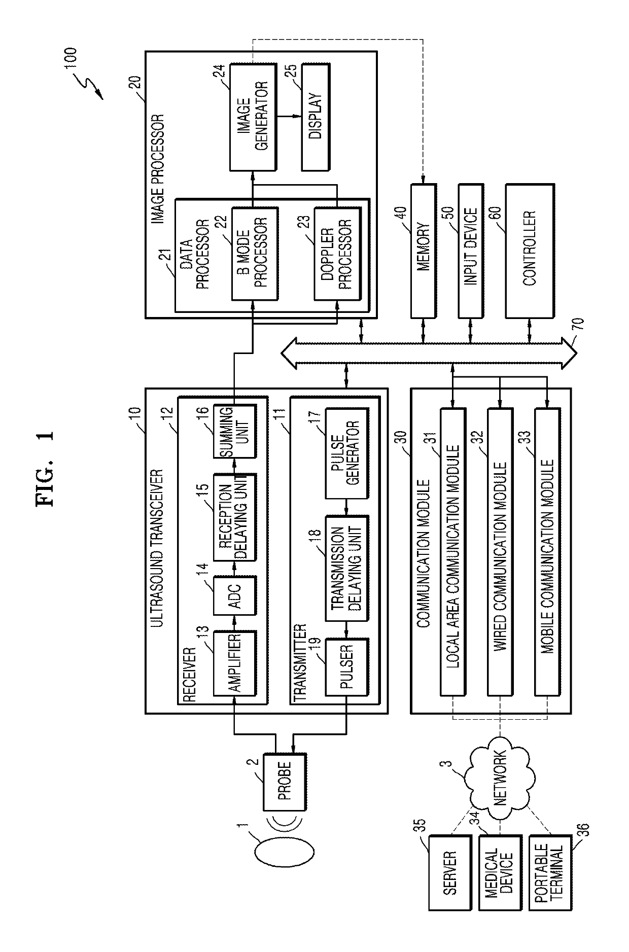
[0009]One or more exemplary embodiments include an ultrasound diagnosis apparatus configured to allow a user to easily and quickly measure biometric parameters.
[0034]According to the one or more exemplary embodiments, the ultrasound diagnosis apparatus enables quicker and easier diagnosis of abnormality of a fetal brain based on three-dimensional (3D) volume data. Furthermore, the ultrasound diagnosis apparatus allows the user to select a desired measurement plane and identify a position of biometric parameters. In addition, inter-observer variance and intra-observer variance in the measurement of an object may be reduced.

Problems solved by technology

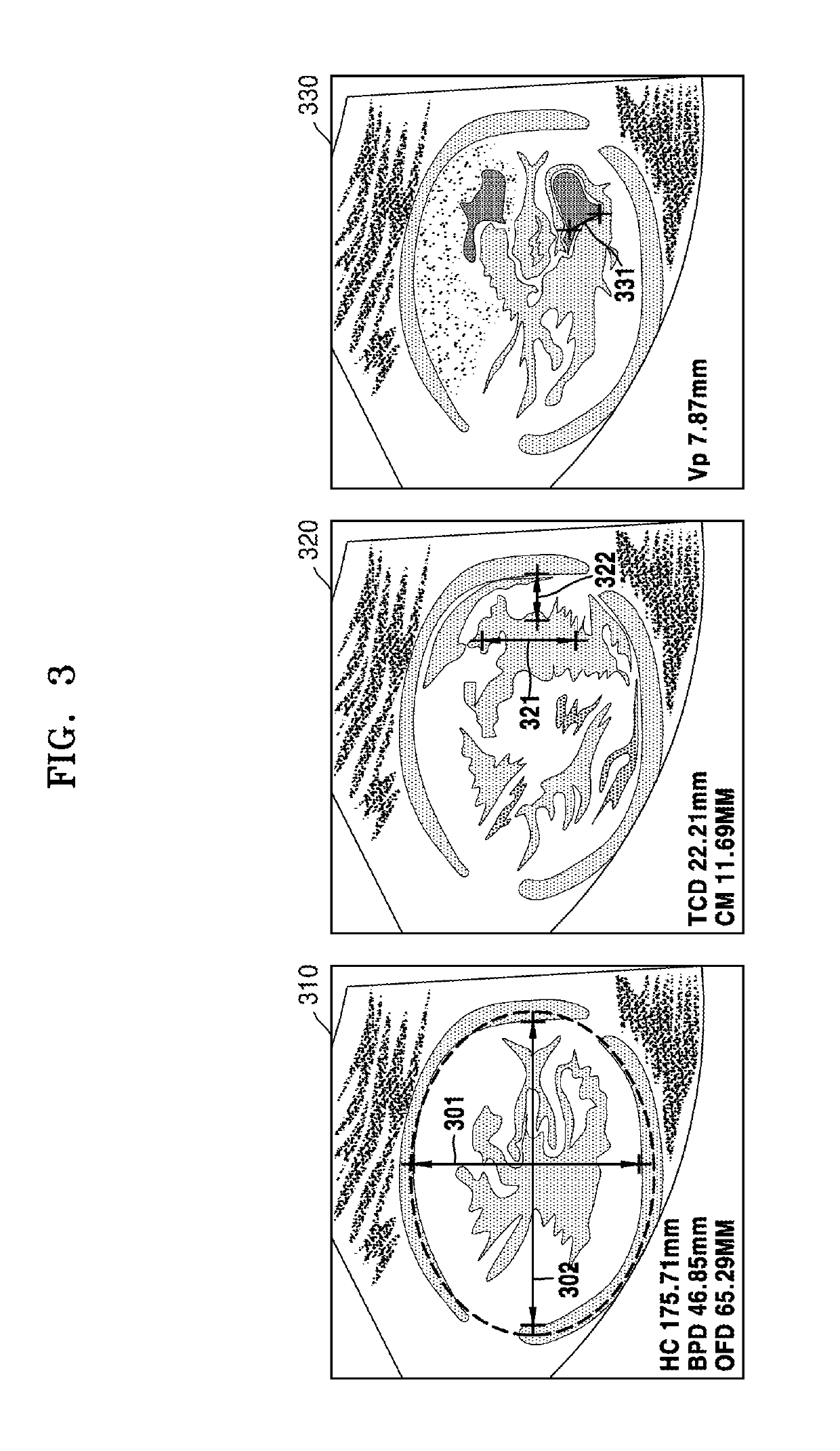
Thus, according to the conventional measuring method, an ultrasound scan needs to be repeated until an ultrasound image of a desired cross-sectional plane is acquired, and thus, it takes a long time to measure a the size of the head of a fetus.
Furthermore, since the accuracy of a cross-sectional plane to be acquired may vary depending on the skill level of a medical practitioner who performs an ultrasound scan, it is difficult to accurately measure fetal HC, OFD, and BPD.

Method used


Image

  • Ultrasound diagnosis apparatus and method for generating image from volume data and displaying the same
  • Ultrasound diagnosis apparatus and method for generating image from volume data and displaying the same
  • Ultrasound diagnosis apparatus and method for generating image from volume data and displaying the same

Examples


Embodiment Construction

[0046]Reference will now be made in detail to exemplary embodiments, examples of which are illustrated in the accompanying drawings, wherein like reference numerals refer to like elements throughout. In this regard, the present exemplary embodiments may have different forms and should not be construed as being limited to the descriptions set forth herein. Accordingly, the exemplary embodiments are merely described below, by referring to the figures, to explain aspects of the present description. Expressions such as “at least one of,” when preceding a list of elements, modify the entire list of elements and do not modify the individual elements of the list.

[0047]The terms used in this specification are those general terms currently widely used in the art in consideration of functions regarding the inventive concept, but the terms may vary according to the intention of those of ordinary skill in the art, precedents, or new technology in the art. Also, some terms may be arbitrarily sel...


PUM


Abstract

Provided is an ultrasound diagnosis apparatus including: a data acquisition unit configured to acquire volume data for a head of an object; an image processor configured to detect a mid-sagittal plane (MSP) from the volume data, generate an MSP image corresponding to the MSP, detect at least one measurement plane based on the MSP, and generate at least one measurement plane image corresponding to the at least one measurement plane; and a display configured to display the MSP image and the at least one measurement plane image on a single screen.

Description

CROSS-REFERENCE TO RELATED APPLICATION[0001]This application claims the benefit of Korean Patent Application No. 10-2014-0100699, filed on Aug. 5, 2014, in the Korean Intellectual Property Office, the disclosure of which is incorporated herein in its entirety by reference.BACKGROUND[0002]1. Field[0003]One or more exemplary embodiments relate to an ultrasound diagnosis apparatus, and more particularly, to an ultrasound diagnosis apparatus for measuring a fetal head based on a three-dimensional (3D) ultrasound image.[0004]2. Description of the Related Art[0005]Ultrasound diagnosis apparatuses transmit ultrasound signals generated by transducers of a probe to an object and receive echo signals reflected from the object, thereby obtaining at least one image of an internal part of the object (e.g., soft tissue or blood flow). In particular, ultrasound diagnosis apparatuses are used for medical purposes including observing an internal area of an object, detecting foreign substances, and a...

Claims


Application Information

Patent Timeline
Patent Type & Authority Patents(United States)
IPC IPC(8): A61B8/08G06T7/62A61B5/00A61B8/00
CPCA61B8/0808A61B8/523A61B5/7267G06T7/62A61B8/0866A61B8/483G06T2207/10136A61B8/468A61B8/5207A61B8/5223A61B8/463G06T2207/30016G06T2207/30044A61B8/461A61B8/13G06T7/0012G06T2207/10132
Inventor LEE, KWANG-HEEKIM, SUNG-YOONYOO, JUN-SANG
Owner SAMSUNG MEDISON CO LTD
Why Eureka
Try Eureka