Method for generating high affinity antibodies
a high affinity antibody and affinity technology, applied in the field of high affinity antibodies, can solve the problem of avoiding the need for time-consuming affinity selection, and achieve the effect of enhancing binding affinity
- Summary
- Abstract
- Description
- Claims
- Application Information
AI Technical Summary
Benefits of technology
Problems solved by technology
Method used
Image
Examples
example 1
n of Micro Beads Coated with a Fast Dissociation Antibody from Micro Beads Coated with a Slow Dissociation Antibody by Flow Cytometry
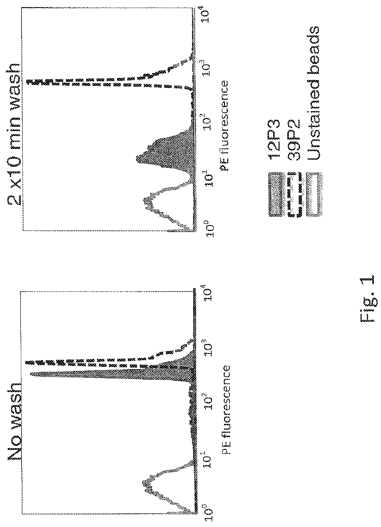
[0063]B cells from immunized mice express BCRs of varying affinity against an immunogen. In an experiment to demonstrate the feasibility of separating B cells by fluorescence-activated cell sorting based on antigen binding dissociation rates using labeled antigen (human ExTek.6His; see Lin, P. et al. PNAS USA. 1998 Jul. 21; 95(15): 8829-8834), polystyrene micro beads (cat. number MPFc-60-5, Spherotech, Lake Forest, Ill.) were coated with mAb 12P3 or 39P2, two mouse monoclonal antibodies against human Tie2 protein. The dissociation t1 / 2 of the binding between these two antibodies and hExTek, a purified protein comprised of human Tie2 extracellular domain and a 6-His tag, is 1.46 min for 12P3 and 198.6 min for 39P2. Polystyrene beads conjugated with polyclonal anti-mFc capture antibodies were incubated with either 12P3 or 39P2 overnight. The next day, th...
example 2
n of Hybridoma Cells Expressing a Low Affinity Antibody from Hybridoma Cells Expressing a High Affinity Antibody by Flow Cytometry
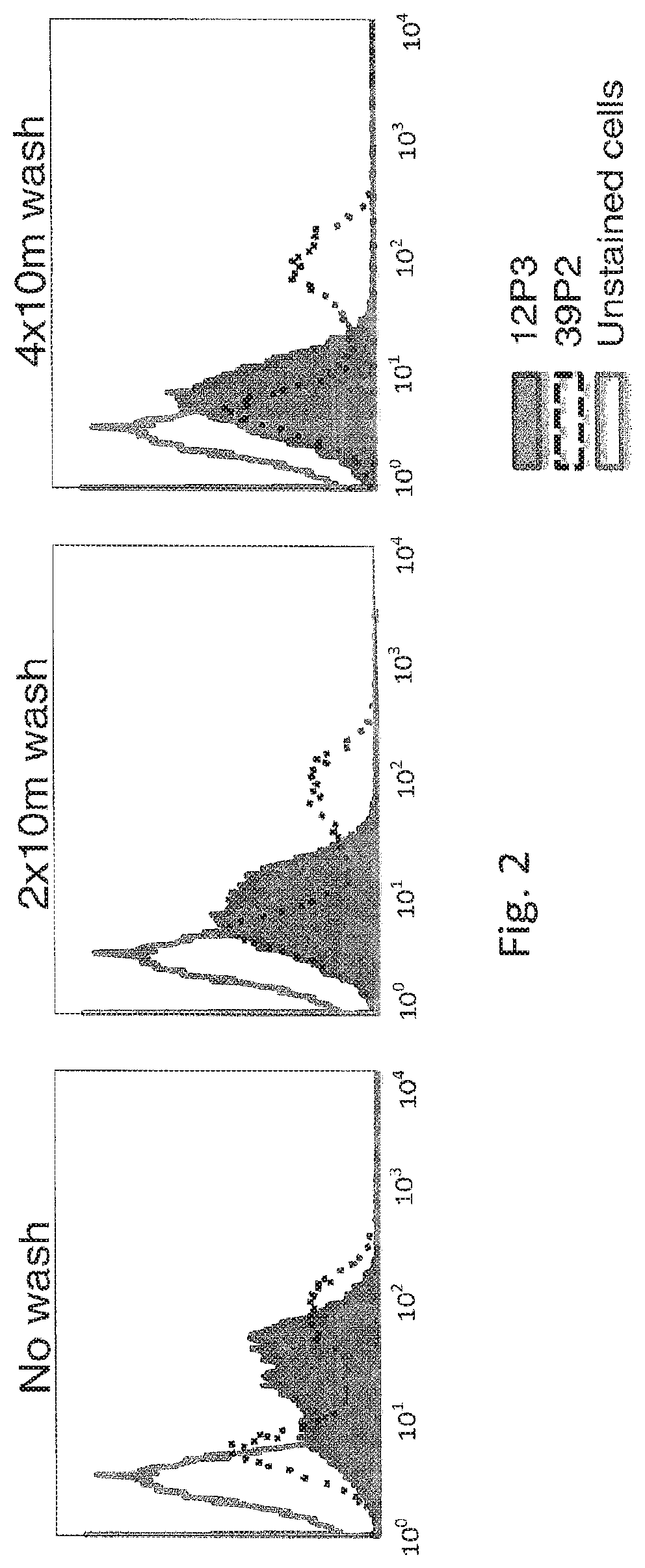
[0066]The hybridoma cell lines producing the 12P3 and 39P2 antibodies expressed membrane-anchored IgG as well as soluble IgG lacking transmembrane domain. The 39P2 cell line contained two populations of cells with different levels of surface IgG expression. These two hybridoma cell lines were stained with 5 nM biotin-hExTek for one hour. The two stained cell populations were each divided into three aliquots. One aliquot for each cell line sample was given excess PE-streptavidin without the removal of biotin-hExTek and incubated for 30 minutes. In parallel, the other aliquots for each cell line sample were washed with excess PBS for either 20 minutes or 40 minutes, then stained with PE-streptavidin for 30 minutes. PE fluorescence of all cell samples was analyzed by FACS (FIG. 2). The 39P2 cell line was comprised of two populations of cells with different l...
example 3
and Sorting Immunized B Cells with Monomeric Antigen Yielded More Antibodies with Slower Dissociate Rate than Staining and Sorting B Cells with Dimer Antigen
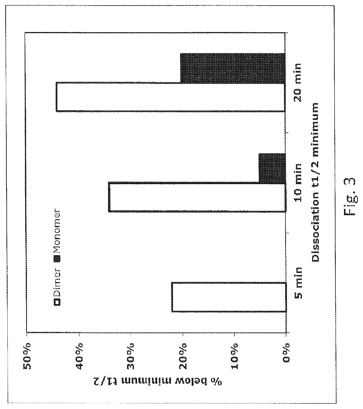
[0068]VELOCIMMUNE® mice were immunized with a purified protein comprised of Tie2 extracellular domain and human Fc domain (Tie2-hFc). This protein is a dimer due to Fc:Fc association. After three injections of Tie2-hFc to boost immune response, mice were sacrificed and splenocytes were harvested. The splenocytes were pooled and divided into two aliquots. Cells in one aliquot were stained with 5 nM biotinylated Tie2-hFc protein and FITC-anti-mFc for 1 hour. In the meantime, the other half of the splenocyte pool was stained with 5 nM biotinylated ExTek and FITC-anti-mFc for 1 hour. The stained cells were washed for 10 minutes with PBS, then stained with SA-PE for one hour. The stained cells were washed once with PBS and were analyzed by flow cytometry on a MoFlo (Cytomation).
[0069]Single IgG positive and antigen positive B cells w...
PUM
| Property | Measurement | Unit |
|---|---|---|
| time | aaaaa | aaaaa |
| dissociation t1/2 | aaaaa | aaaaa |
| dissociation t1/2 | aaaaa | aaaaa |
Abstract
Description
Claims
Application Information
 - R&D
- Intellectual Property
- Life Sciences
- Materials
- Tech Scout
- Unparalleled Data Quality
- Higher Quality Content
- 60% Fewer Hallucinations
Browse by: Latest US Patents, China's latest patents, Technical Efficacy Thesaurus, Application Domain, Technology Topic, Popular Technical Reports.
© 2025 PatSnap. All rights reserved.Legal|Privacy policy|Modern Slavery Act Transparency Statement|Sitemap|About US| Contact US: help@patsnap.com


