

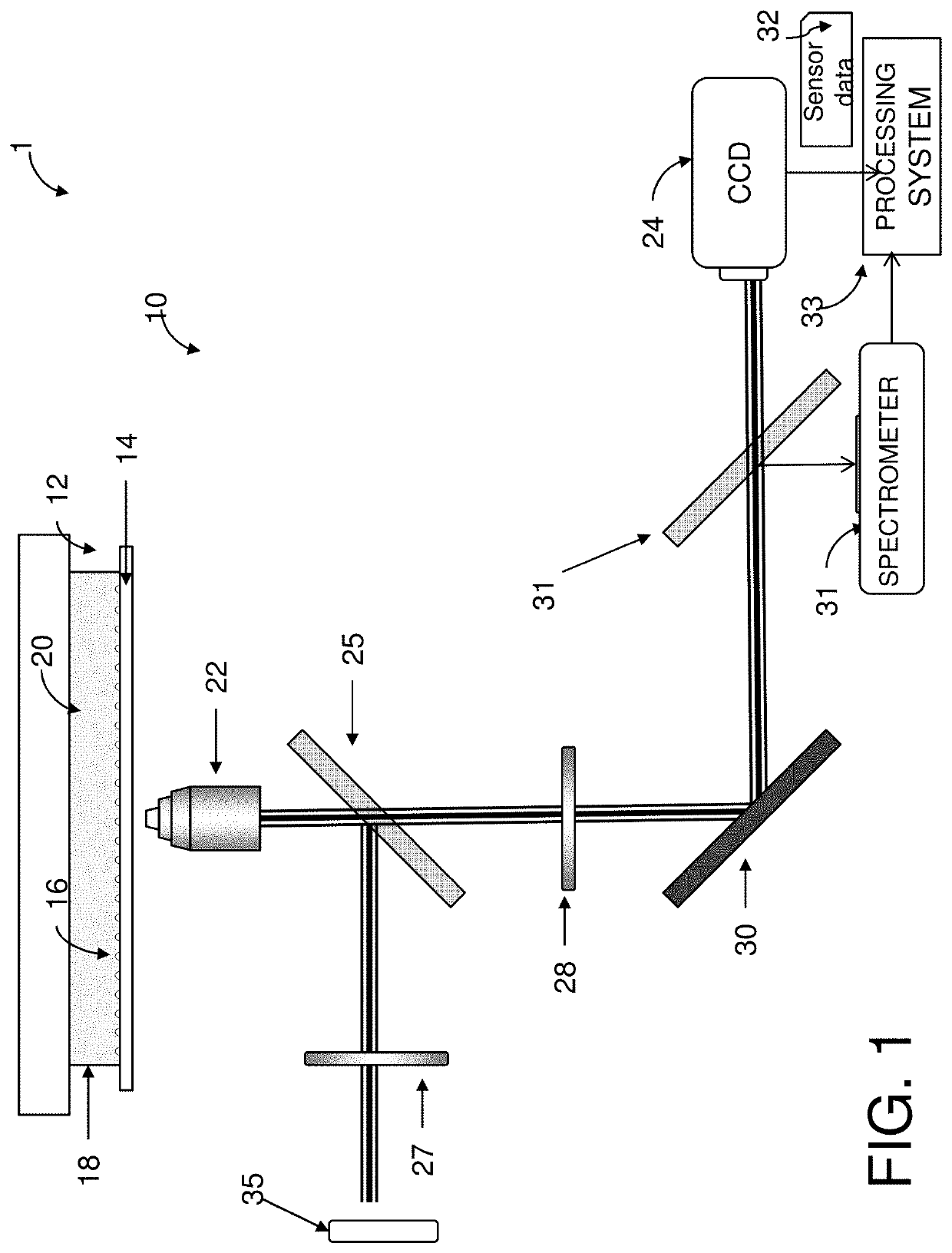
Determining extracellular analyte concentration with nanoplasmonic sensors
a nano-plasmonic sensor and analyte technology, applied in the direction of fluorescence/phosphorescence, instruments, analyte processes for specific applications, etc., can solve the problems of limiting the use of techniques, requiring invasiveness, and unable to measure extracellular protein
- Summary
- Abstract
- Description
- Claims
- Application Information
AI Technical Summary
Problems solved by technology
Method used
Image

Examples
examples
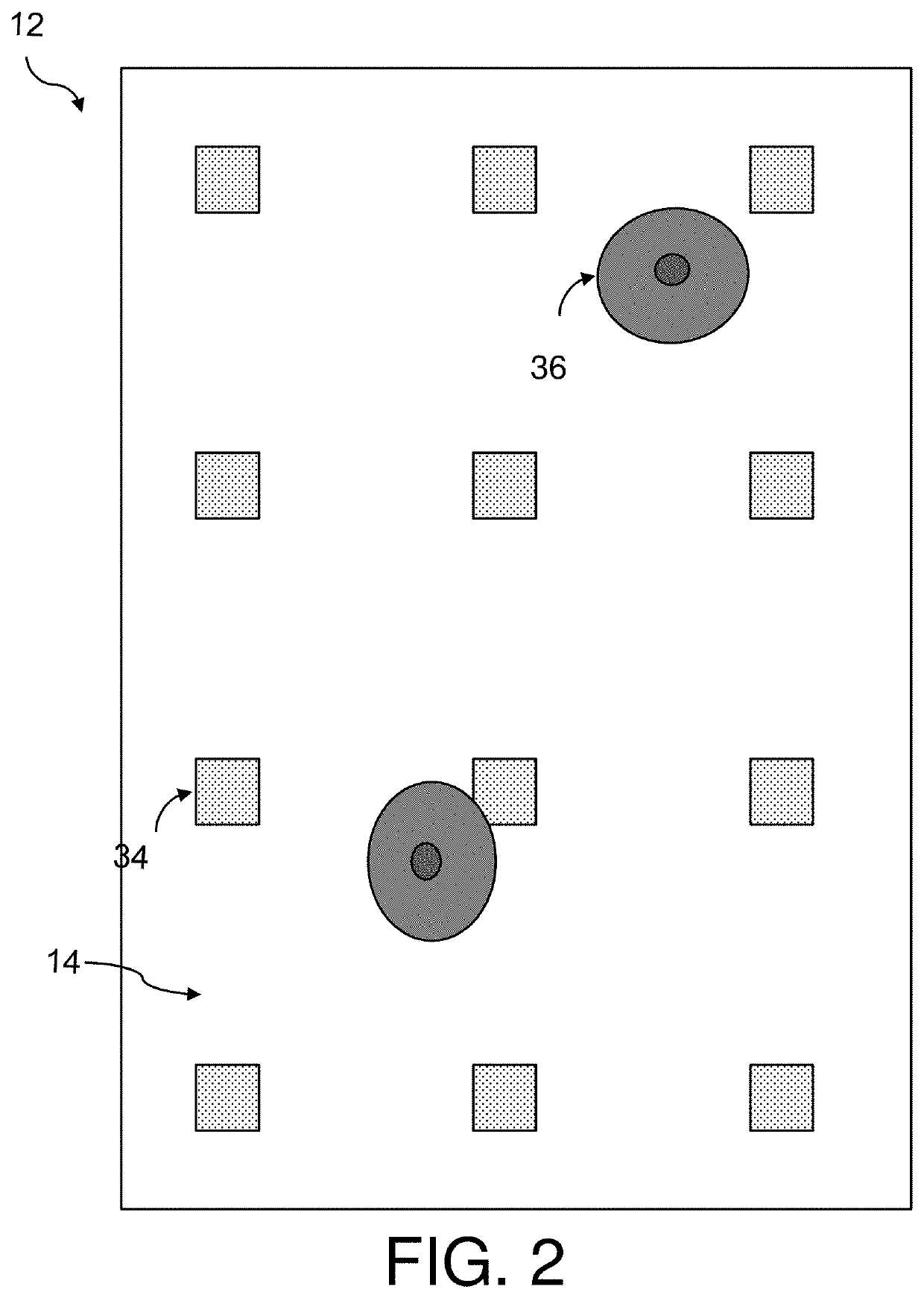
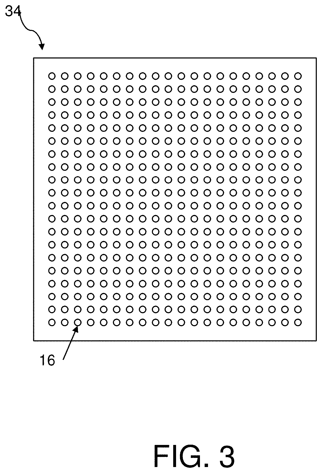
1. Fabrication and Functionalization of Plasmonic Nanostructures
[0138]Arrays of nanostructures were patterned onto No. 1.5 glass coverslips by spinning a bilayer resist structure consisting of polymethyl methacrylate and ethyl lactate methyl methacrylate copolymer with thicknesses of 180 nm and 250 nm respectively. The resist was electron-beam patterned using doses of 300 μC / cm2 and subsequently developed for one minute in a 2:1 solution of isopropyl alcohol:methyl isobutyl ketone. A 5 nm layer of Ti followed by 70 nm of Au was deposited with a Temescal electron-beam evaporator. The bilayer resist was then lifted off by soaking in acetone for 4 hours.
[0139]Radio frequency (RF) plasma ashing (40 W) with 300 mTorr of a 5% hydrogen, 95% argon mixture was used to clean the glass and gold surfaces on the chips. The gold nanostructures were functionalized in a two-component ethanolic-based thiol bath (0.5 mM), containing a 3:1 ratio of SH—(CH2)8-EG3-OH to SH—(CH2)11-EG3-NH2 for 18 hours, ...
PUM
| Property | Measurement | Unit |
|---|---|---|
| height | aaaaa | aaaaa |
| diameter | aaaaa | aaaaa |
| diameter | aaaaa | aaaaa |
Abstract
Description
Claims
Application Information