Inhibitors of extracellular Hsp90

Active Publication Date: 2006-04-06
TUFTS UNIV
View PDF5 Cites 35 Cited by
  • Summary
  • Abstract
  • Description
  • Claims
  • Application Information

AI Technical Summary

Benefits of technology

[0036] The term “biogonjugate” as used herein means that an inhibitor of extracellular Hsp90 is covalently or non-covalently linked and/or coupled to or with, respectively, another protein, a solid matrix (e.g. like a bead), wit

Problems solved by technology

While Hsp90 has well-established functions intracellularly, reports of extracellular occurrence and its function are scarce.
Inhibitors such as the geldanamycin analogue 17-AAG are already being tested in clinical trials but there are concerns about toxicity resulting from non-specific inhibition

Method used


Image

  • Inhibitors of extracellular Hsp90
  • Inhibitors of extracellular Hsp90
  • Inhibitors of extracellular Hsp90

Examples


example 1

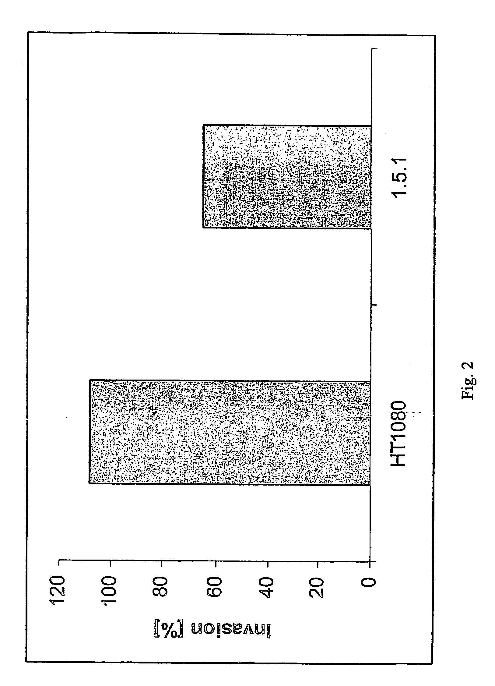
Generation of Monoclonal Antibodies (e.g. mAb1.5.1)

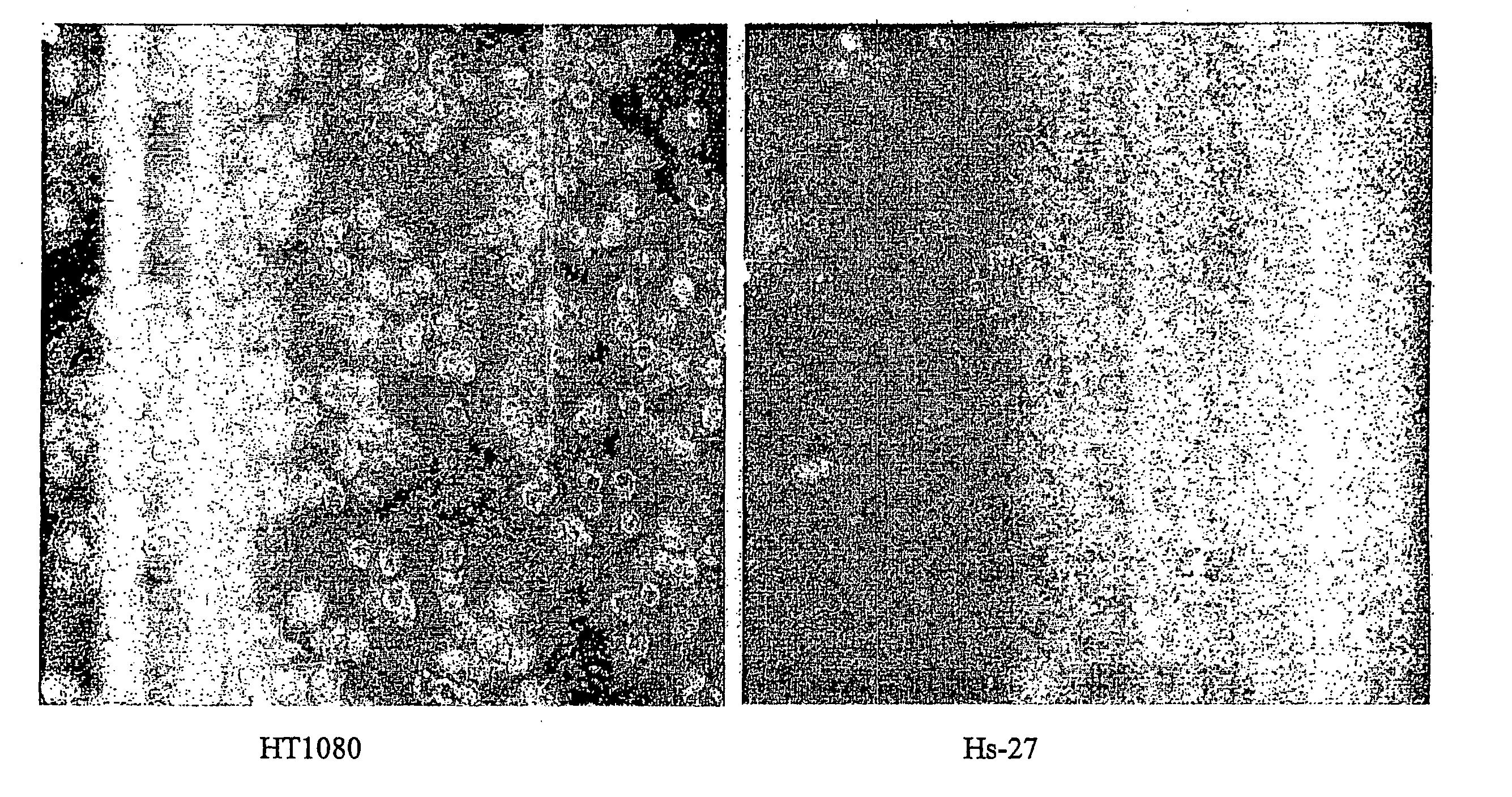
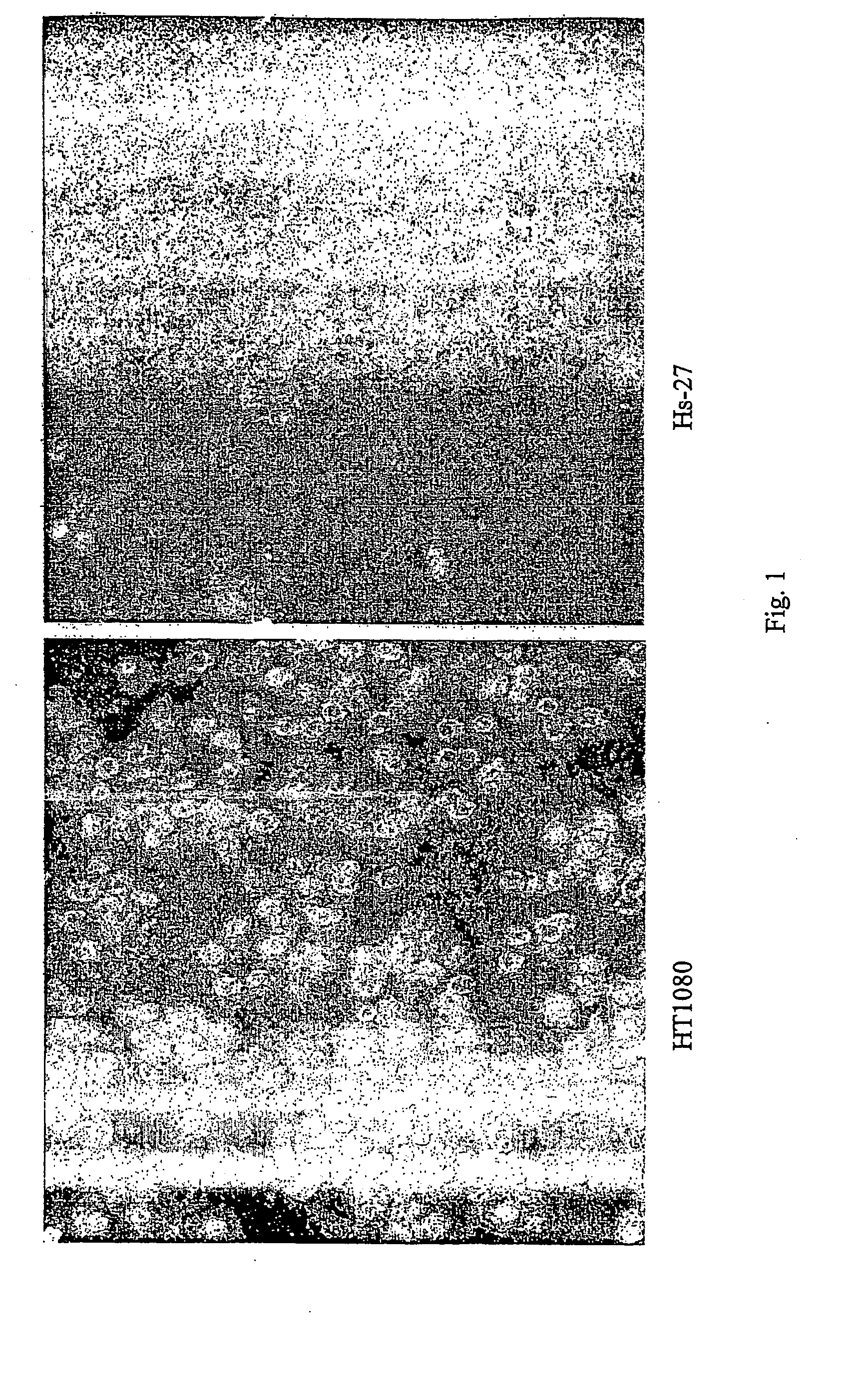
[0155] Mice were immunized with either fixed or lysed HT-1080 cells twice over three months. For fixation, confluent (1×10⁷ / 75 cm2 flask) HT-1080 cells were washed once with PBS, then removed with PBS and scraping. Cells were resuspended by pipeting, centrifuged for 5 minutes at 220×g, and fixed with 10 mL of 2% paraformaldehyde for 10 minutes at 4° C. Cells were washed with PBS, then resuspended in 1 ML of PBS. Lysed cells were generated by trypsinizing confluent HT-1080 cells, washing once with PBS, then pelleting for 1 minute at 12000×g. Cells were then lysed in lysis buffer (0.67 Triton-X-100 in 0.33M Tris-HCl, pH 7.5) at room temperature for 5 minutes. Lysate was then centrifuged for 5 minutes at 12000×g. Triton-X-100 was removed by adding the lysate to PD-10 columns and eluting protein with 3.5 mL PBS. Protein was quantitated using Micro-BCA kit (Pierce), and 150 μg / mL / mouse was used for immunization.

[0156] Generation of hyb...

example 2

Construction of an Immune Library

[0158] Two BALB / c mice were each immunized intradermally with 2×10⁷ paraformaldehyde fixed HT-1080 cells (human fibrosarcoma cell line; ATCC, CCL-121). Following the first immunization, the injections were repeated twice in a period of 39 days, the mice sacrificed and the spleens isolated and frozen in liquid nitrogen.

[0159] Total RNA was isolated using the RNeasy Midi Kit (QIAGEN #75142) as described by the manufacturer using half of each spleen preparation. The RNA concentration and purity was determined by a denaturing formaldehyde gel and photometric measurement.

[0160] cDNA was synthesized using 8.9 μg of freshly prepared RNA and 10 pmol of a primer mix IgG1-c, IgG2a-c, IgG2b-c, IgG3-c, VLL-c, VLK-c) using the Superscript™ II Kit (GibcoBRL Life Technologies #18064-014). These primers anneal to the RNA encoding the IgG heavy-chain (VH) genes and the light chain (VL) genes of the kappa and lambda family. VH genes were PCR amplified from 1 μl of ...

example 3

Selection and Screening of scFv (Selection on Fixed Cells)

[0161] Single chain Fv were selected from a phage display library generated from mice immunized with fixed HT-1080 cells. The library was generated using the phage display vector pXP10.

[0162] Phages expressing scFv with high affinity to tumor cells were selected as follows: HT-1080 cells were harvested with 0.05% EDTA, fixed with paraformaldehyde, diluted to 1×10⁷ cells / ml in PBS and immobilized onto wells of a 96 well UV cross-link plate (Corning Costar). The wells of the UV cross-link plate were blocked with 5% Skim Milk Powder (#70166, Fluka) in PBS (MPBS). 1012 cfu (colony forming units) of phage library / 106 cells were pre-blocked for 1 hour at 25° C. with MPBS and subsequently incubated for 1.5 hour at room temperature (RT) with the cells. The wells of the UV cross-link plate were washed six times with PBS+0.05% Tween-20 followed by six washes with PBS. Bound phage were eluted by the addition of 10 mM Glycine pH 2.2, a...


PUM


Abstract

The present invention describes inhibitors of extracellular Hsp90. The inhibition of extracellular Hsp90 leads to a reduction of the invasiveness of the tumor cells. Furthermore, the invention relates to the use of molecules inhibiting extracellular Hsp90 function for the manufacture of a medicament for the treatment or prevention of invasion and/or metastatic potential of cancer cells.

Description

[0001] This invention was made with government support under CA81668 awarded by the National Institutes of Health. The government has certain rights in the invention.BACKGROUND OF THE INVENTION [0002] Malignant tumors shed cells, which migrate to new tissues and create secondary tumors. The process of generating secondary tumors is called metastasis and is a complex process in which tumor cells colonize sites distant from the primary tumor. Liotta ((1986) Cancer Res. 46, 1-7) has proposed a three-step hypothesis for the process of metastasis: The first step is tumor cell attachment via cell surface receptors. The anchored tumor cell next secretes hydrolytic enzymes or induces host cells to secrete enzymes, which can degrade the matrix locally. Matrix lysis most likely takes place in a highly localized region close to the tumor cell surface. The third step is tumor cell locomotion into the region of the matrix modified by proteolysis. Thus, invasion of the matrix is not merely due to...

Claims


Application Information

Patent Timeline
IPC IPC(8): A61K39/395A61K48/00A61K31/366
CPCA61K31/366A61K2039/505C07K16/30C07K2317/622A61K39/39558G01N33/57484A61K31/395A61K47/4823G01N33/574A61K47/48923C07K2317/34A61K47/6939A61P35/04
Inventor JAY, DANIELEUSTACE, BRENDASAKURAI, TAKASHI
Owner TUFTS UNIV
Why Eureka
Try Eureka