Isolated peptides which bind to MHC class II molecules, and uses thereof
a technology of mhc class ii and peptides, which is applied in the field of isolated peptides which bind to mhc class ii molecules, can solve the problems of inability to know the exact method of purification, inability to purify cells, and inability to meet the requirements of a single peptide,
- Summary
- Abstract
- Description
- Claims
- Application Information
AI Technical Summary
Problems solved by technology
Method used
Image
Examples
example 2
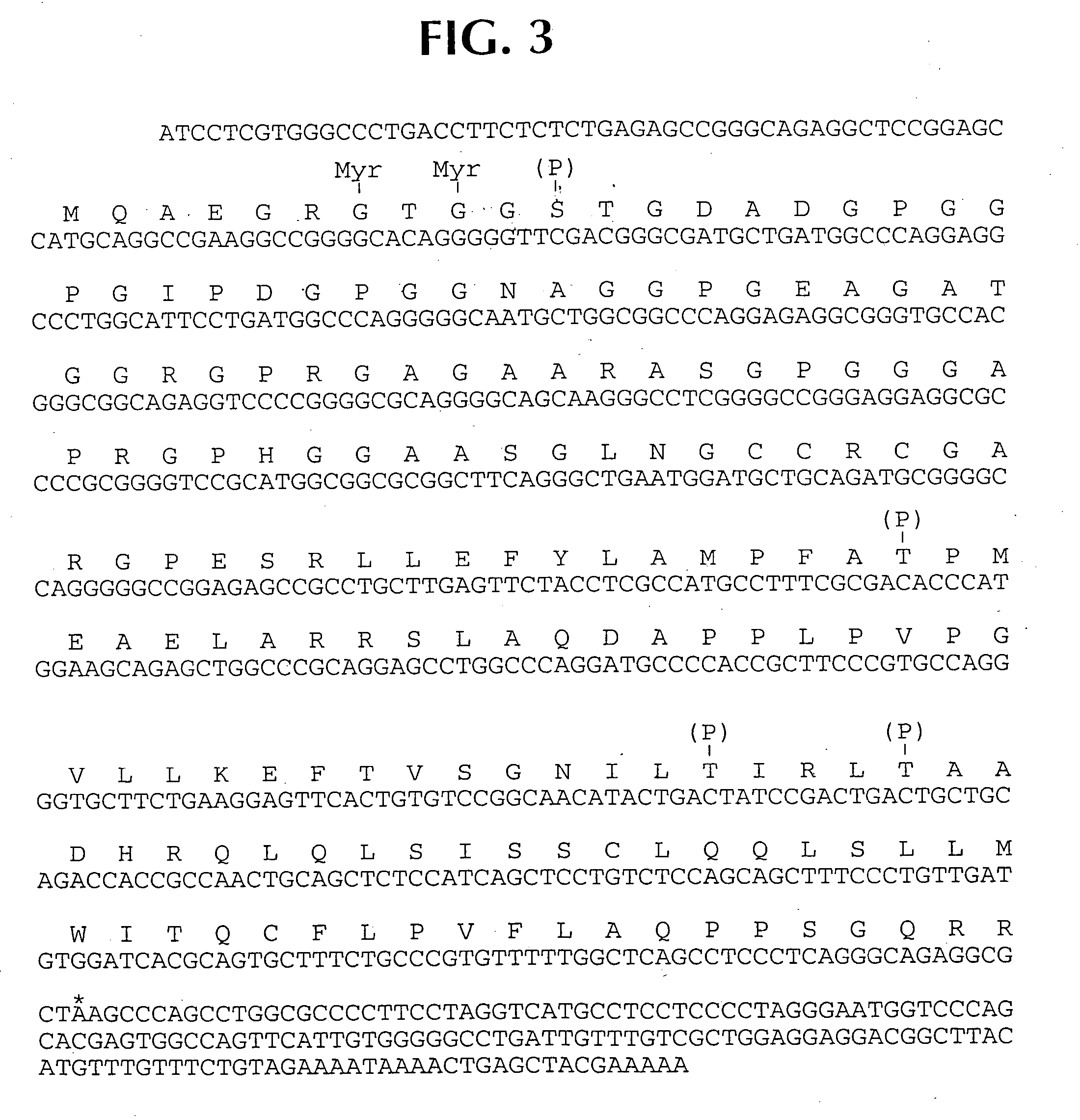
[0024] Following identification, the reactive clones were subcloned to monoclonality via dilution cloning and testing with human serum. These clones were then purified, excised in vitro, and converted into pBK-CMV plasmid forms, using the manufacturer's instructions. The inserted DNA was then evaluated using EcoRI-XbaI restriction mapping to determine different inserts. Eight different inserts were identified, ranging in size from about 500 to about 1.3 kilobase pairs. The clones were sequenced using an ABI PRISM automated sequencer.
[0025] Table 1 summarizes the results. One gene was represented by four overlapping clones, a second by three overlapping clones, and the remaining six by one clone only.
[0026] A homology search revealed that the clones referred to as NY-ESO-2, 3, 6, 7 were already known. See Elisei, et al., J. Endocrin. Invest. 16: 533-540 (1993); Spritz, et al., Nucl. Acids Res. 15: 10373-10391 (1987); Rabbits, et al., Nature Genetics 4: 175-180 (1993); Crozat, et al.,...
example 3
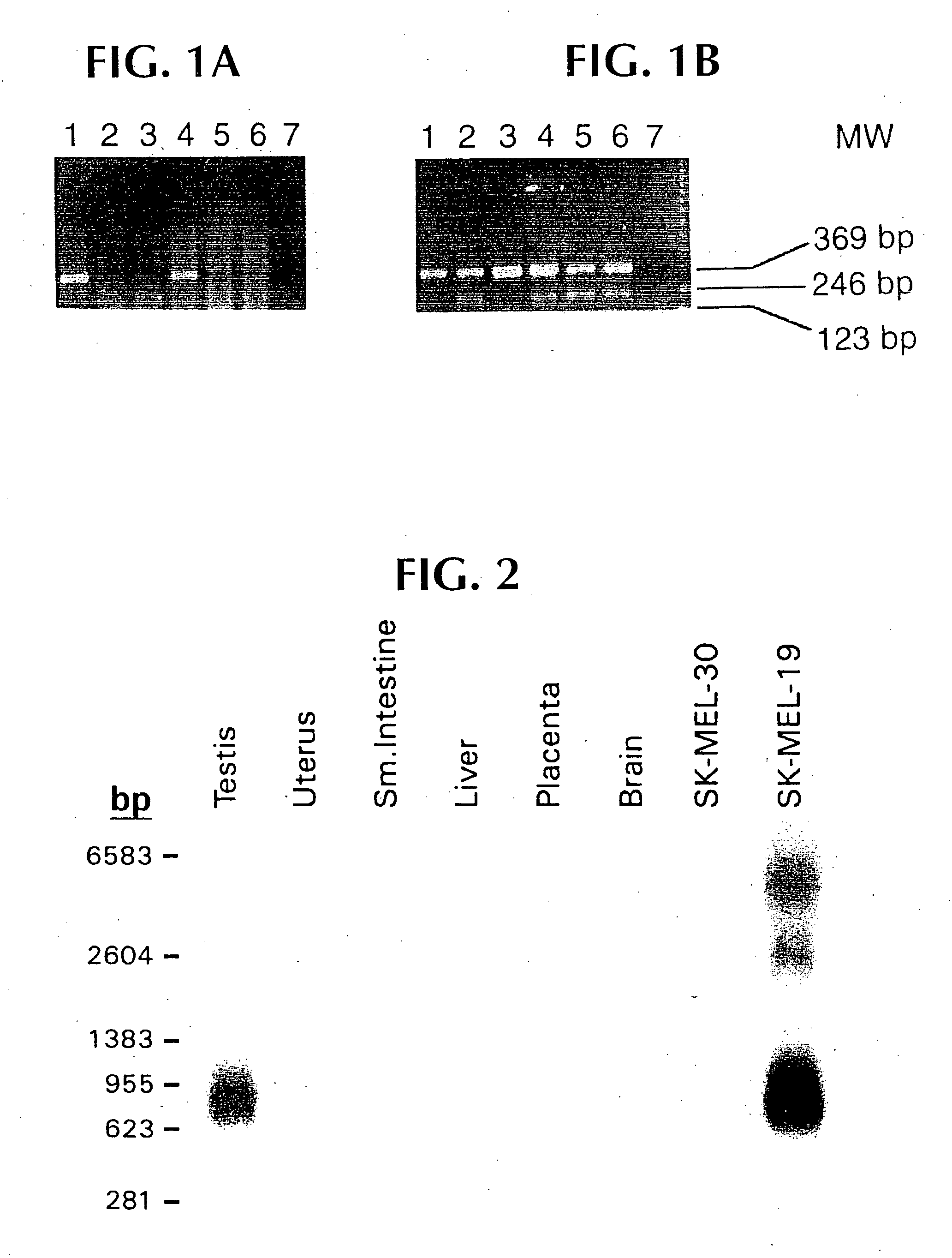
[0027] Studies were carried out to evaluate mRNA expression of the NY-ESO 1, 4, 5 and 8 clones. To do this, specific oligonucleotide primers were designed for each sequence, such that cDNA segments of 300-400 base pairs could be amplified, and so that the primer melting temperature would be in the range of 65-70.degree. C. Reverse transcription-PCR was then carried out using commercially available materials and standard protocols. A variety of normal and tumor cell types were tested. The clones NY-ESO-4 and NY-ESO-8 were ubiquitous, and were not studied further. NY-ESO-5 showed high level expression in the original tumor, and in normal esophageal tissue, suggesting that it was a differentiation marker.
[0028] NY-ESO-1 was found to be expressed in tumor mRNA and in testis, but not normal colon, kidney, liver or brain tissue. This pattern of expression is consistent with other tumor rejection antigen precursors.
example 4
[0029] The RT-PCR assay set forth supra was carried out for NY-ESO-1 over a much more complete set of normal and tumor tissues. Tables 2, 3 and 4 show these results. In brief, NY-ESO-1 was found to be highly expressed in normal testis and ovary cells. Small amounts of RT-PCR production were found in normal uterine myometrium, and not endometrium, but the positive showing was not consistent. Squamous epithelium of various cell types, including normal esophagus and skin, were also negative.
[0030] When tumors of unrelated cell lineage were tested, 2 of 11 melanomas cell lines showed strong expression, as did 16 of 67 melanoma specimens, 6 of 33 breast cancer specimens and 4 of 4 bladder cancer. There was sporadic expression in other tumor types.
2TABLE 2 mRNA distribution of NY-ESO-1 in normal tissues Tissue mRNA Tissue mRNA Esophagus -Adrenal - Brain* - Pancreas - Fetal Brain - Seminal Vesicle - Heart - Placenta - Lung - Thymus -Liver - Lymph node - Spleen - Tonsil - Kidney - PBL -Stom...
PUM
| Property | Measurement | Unit |
|---|---|---|
| melting temperature | aaaaa | aaaaa |
| concentration | aaaaa | aaaaa |
| molecular mass | aaaaa | aaaaa |
Abstract
Description
Claims
Application Information
 - R&D
- Intellectual Property
- Life Sciences
- Materials
- Tech Scout
- Unparalleled Data Quality
- Higher Quality Content
- 60% Fewer Hallucinations
Browse by: Latest US Patents, China's latest patents, Technical Efficacy Thesaurus, Application Domain, Technology Topic, Popular Technical Reports.
© 2025 PatSnap. All rights reserved.Legal|Privacy policy|Modern Slavery Act Transparency Statement|Sitemap|About US| Contact US: help@patsnap.com


