Automatic optimal view determination for cardiac acquisitions

a technology of optimal view and cardiac image, applied in the field of medical imaging, can solve problems such as not always yielding an appropriate coordinate fram

Inactive Publication Date: 2005-02-10
SIEMENS MEDICAL SOLUTIONS USA INC
View PDF5 Cites 26 Cited by
  • Summary
  • Abstract
  • Description
  • Claims
  • Application Information

AI Technical Summary

Benefits of technology

[0007] An exemplary embodiment of the present invention includes a method of determining optimal viewing planes for a cardiac image acquisition. The method includes acquiring a set of sagittal, axial, and coronal images of a heart, where the axial and coronal images intersect with the sagittal image orthogonally, and where the heart has a natural axis and a left ventricle (“LV”) with a bloodpool, a bloodpool border, and an apex. The method also includes making a map of the bloodpool border, and using the map to create a full coordinate frame oriented along the natural axis.
[0008] Another exemplary embodiment of the present invention includes a system for determi

Problems solved by technology

Since ventricular shapes can vary, some even being banana-shaped,

Method used


Image

  • Automatic optimal view determination for cardiac acquisitions
  • Automatic optimal view determination for cardiac acquisitions
  • Automatic optimal view determination for cardiac acquisitions

Examples


Embodiment Construction

[0021] Exemplary embodiments of the present invention provide methods, systems, and apparatus for determining the optimal short axis and long axis viewing planes for cardiac image acquisitions. The images can be acquired using: a Magnetic Resonance Scanner (“MR”), a Positron Emission Tomography Scanner (“PET”), a Single Photon Emission Computed Tomography (“SPECT”), a Computed Tomography Scanner (“CT”), and other medical imaging devices. CT, SPECT, and PET volume data of a heart, among other data sources representative of a heart, can be reformatted, subsequent to acquisition, to create the desired images as well. After the optimal viewing planes have been determined, the images can be rescanned or the data, like that of CT volumes, can be reformatted to acquire new images at the new viewing planes.

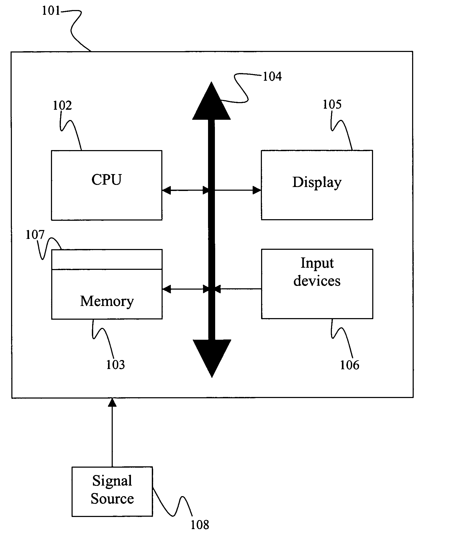
[0022] Referring to FIG. 1, according to an exemplary embodiment of the present invention, a computer system 101 for implementing the present invention includes a central processing unit...


PUM


Abstract

A method, system, and apparatus of determining optimal viewing planes for cardiac image acquisition, wherein the method includes acquiring a set of sagittal, axial, and coronal images of a heart, where the axial and coronal images intersect with the sagittal image orthogonally, and where the heart has a natural axis and a left ventricle (“LV”) with a bloodpool, a bloodpool border, and an apex. The method also includes making a map of the bloodpool border, and using the map to create a full coordinate frame oriented along the natural axis.

Description

CROSS-REFERENCE TO RELATED APPLICATION [0001] This application claims the benefit of U.S. Provisional Application Ser. No. 60 / 473,730 (Attorney Docket No. 2003P07843US), filed on 28 May 2003 and entitled “Automatic Optimal View Determination for Cardiac Acquisitions”, which is incorporated herein by reference in its entirety.BACKGROUND OF INVENTION [0002] 1. Technical Field [0003] The present invention relates to medical imaging, and more particularly, to determining the short and long axis viewing planes for cardiac image acquisitions. [0004] 2. Discussion of the Related Art [0005] In the field of medical imaging, images oriented around the short and long axis normals of the heart are the standard format for evaluation by clinicians. The orientation of the heart, and therefore its short and long axis normals, are unique to individuals. Thus, in acquiring such images, the orientation of the individual's heart and its associated coordinate frame (short axis, long axis, and the direct...

Claims


Application Information

Patent Timeline
IPC IPC(8): G06T5/00G06T7/00
CPCG06T7/004G06T2207/30048G06T2207/10081G06T7/0083G06T7/12G06T7/70
Inventor O'DONNELL, THOMASCOWAN, BRETTYOUNG, ALISTAIR
Owner SIEMENS MEDICAL SOLUTIONS USA INC
Why Eureka
Try Eureka