
Transmural ablation device with parallel electrodes

a transmural ablation and parallel electrode technology, applied in the field of transmural ablation devices with parallel electrodes, can solve the problems of rf energy or ultra-low temperature freezing to the inside of the heart chamber, difficult positioning of catheters and associated electrodes or probes, and difficulty in precisely locating aberrant tissue, so as to reduce the likelihood of thrombosis

Inactive Publication Date: 2005-02-10
ATRICURE
View PDF99 Cites 97 Cited by
  • Summary
  • Abstract
  • Description
  • Claims
  • Application Information

AI Technical Summary

Benefits of technology

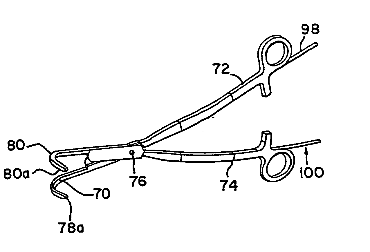
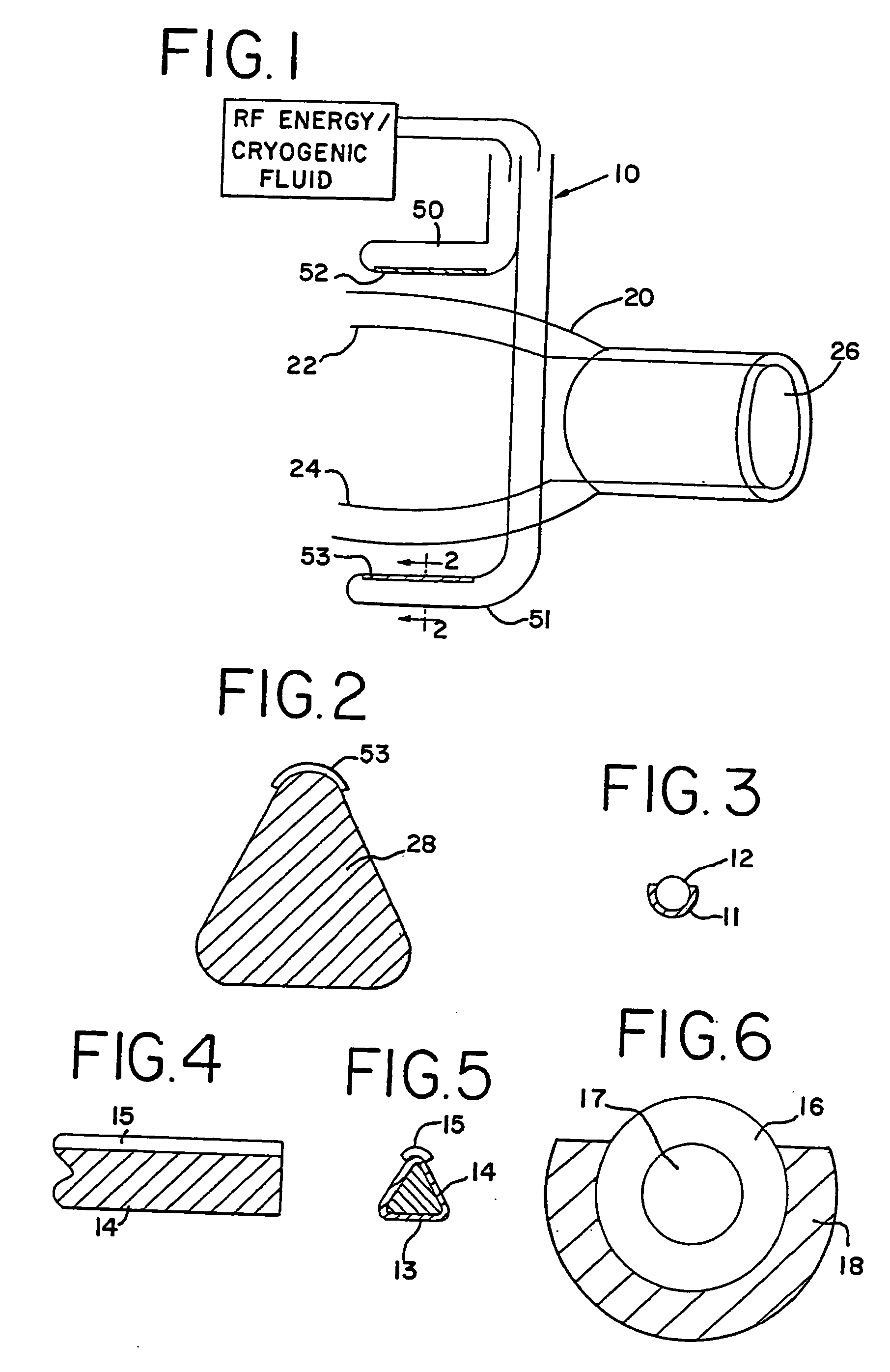
[0022] These objects, and others which will become apparent upon reference to the following detailed description and attached drawings, are achieved by the use of a clamping and ablating device for use in treating cardiac arrhythmia having first and second handle members, with first and second mating parallel jaw members associated with the first and second handle members, respectively. The jaw members are preferably curved and are movable by the handle members between a first open position and a second clamped position, and the jaw members have insulated outer surfaces which may have convex, opposed mating surfaces. Each mating surface has a central region, with the central region of the first jaw being aligned with the central region of the second jaw. A first elongated electrode extends along the central region of the first jaw and a second elongated electrode extends along the central region of the second jaw. The first and second electrodes are adapted to be connected to an RF energy source so that, when activated, the electrodes are of opposite polarity. In a preferred embodiment, the electrodes are made of gold-plated copper and measure between approximately 3 to 8 cm in length and approximately 0.12 to 0.6 mm in width. By the use of such a device a clamping zone is created that is approximately at least three times wider than the contact zone of the electrodes with the tissue. This permits the ablation to be performed with a minimum of contact between the electrodes and any blood cells, thus greatly reducing the likelihood of thrombus. The design also allows for a minimum distance between the electrodes, further encouraging complete, transmural ablation that creates a continuous lesion in a single step.

Problems solved by technology

Common problems encountered in this procedure are difficulty in precisely locating the aberrant tissue, and complications related to the ablation of the tissue.
Locating the area of tissue causing the arrhythmia often involves several hours of electrically “mapping” the inner surface of the heart using a variety of mapping catheters, and once the aberrant tissue is located, it is often difficult to position the catheter and the associated electrode or probe so that it is in contact with the desired tissue.
The application of either RF energy or ultra-low temperature freezing to the inside of the heart chamber also carries several risks and difficulties.
It is very difficult to determine how much of the catheter electrode or cryogenic probe surface is in contact with the tissue since catheter electrodes and probes are cylindrical and the heart tissue cannot be visualized clearly with existing fluoroscopic technology.
Further, because of the cylindrical shape, some of the exposed electrode or probe area will almost always be in contact with blood circulating in the heart, giving rise to a risk of clot formation.
Clot formation is almost always associated with RF energy or cryogenic delivery inside the heart because it is difficult to prevent the blood from being exposed to the electrode or probe surface.
Some of the RF current flows through the blood between the electrode and the heart tissue and this blood is coagulated, or frozen when a cryogenic probe is used, possibly resulting in clot formation.
When RF energy is applied, the temperature of the electrode is typically monitored so as to not exceed a preset level, but temperatures necessary to achieve tissue ablation almost always result in blood coagulum forming on the electrode.
Overheating or overcooling of tissue is also a major complication, because the temperature monitoring only gives the temperature of the electrode or probe, which is, respectively, being cooled or warmed on the outside by blood flow.
The actual temperature of the tissue being ablated by the electrode or probe is usually considerably higher or lower than the electrode or probe temperature, and this can result in overheating, or even charring, of the tissue in the case of an RF electrode, or freezing of too much tissue by a cryogenic probe.
Overheated or charred tissue can act as a locus for thrombus and clot formation, and over freezing can destroy more tissue than necessary.
It is also very difficult to achieve ablation of tissue deep within the heart wall.
As the depth of penetration increases, the time, power, and temperature requirements increase, thus increasing the risk of thrombus formation.
Multielectrode catheters have been developed which can be left in place, but continuity can still be difficult to achieve, and the lesions created can be quite wide.
Because of the risks of char and thrombus formation, RF energy, or any form of endocardial ablation, is rarely used on the left side of the heart, where a clot could cause a serious problem (e.g., stroke).
Because of the physiology of the heart, it is also difficult to access certain areas of the left atrium via an endocardial, catheter-based approach.
However, it is still difficult to create long, continuous lesions, and it is difficult to achieve good depth of penetration without creating a large area of ablated tissue.
When used from an endocardial approach, the limitations of all energy-based ablation technologies to date are the difficulty in achieving continuous transmural lesions, and minimizing unnecessary damage to endocardial tissue.
However, this technology creates rather wide (greater than 5 mm) lesions which could lead to stenosis (narrowing) of the pulmonary veins.
Additionally, there is no feedback to determine when full transmural ablation has been achieved.
Consequently, there is no energy delivered at the backplate / tissue interface intended to ablate tissue.
It is important to note that all endocardial ablation devices that attempt to ablate tissue through the full thickness of the cardiac wall have a risk associated with damaging structures within or on the outer surface of the cardiac wall.
As an example, if a catheter is delivering energy from the inside of the atrium to the outside, and a coronary artery, the esophagus, or other critical structure is in contact with the atrial wall, the structure can be damaged by the transfer of energy from within the heart to the structure.
The coronary arteries, esophagus, aorta, pulmonary veins, and pulmonary artery are all structures that are in contact with the outer wall of the atrium, and could be damaged by energy transmitted through the atrial wall.

Method used


Image

  • Transmural ablation device with parallel electrodes
  • Transmural ablation device with parallel electrodes
  • Transmural ablation device with parallel electrodes

Examples


Embodiment Construction

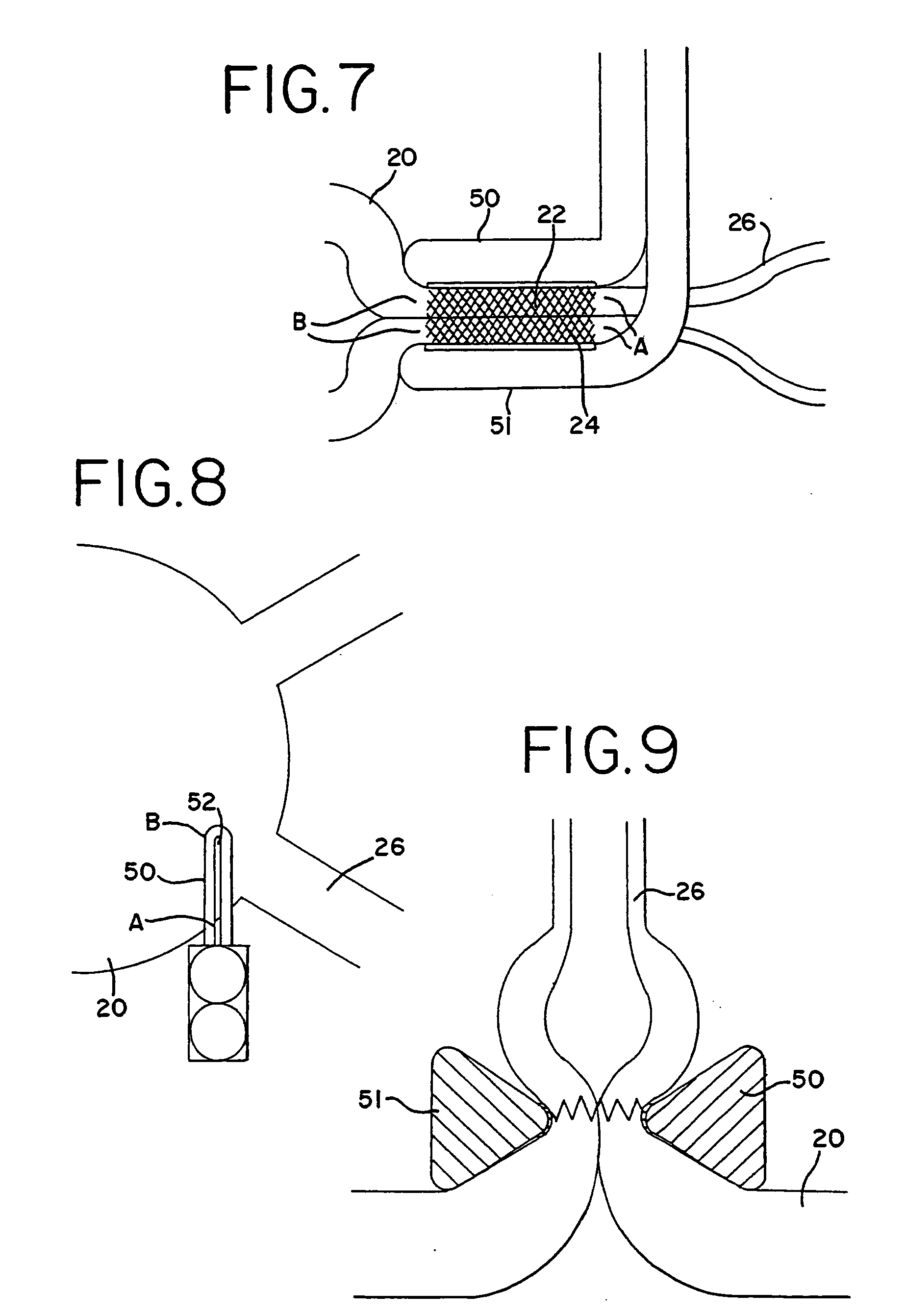
[0095] With reference to the present invention, the compression of the atrial tissue is important because it insures that the exposed electrode surface or cryogenic probe is not in contact with any tissue or blood except the clamped tissue to be ablated. Specifically, the clamping of the tissue between the electrodes or cryogenic probes insures that the conductive or cooled area is only in contact with the clamped tissue. The compressed tissue acts to isolate the electrically active or cryogenically cooled surface, and prevents inadvertent energy delivery to other parts of the heart or blood. The outside temperature of the electrode can easily be monitored to insure that the temperature of the insulation in contact with blood remains below a critical temperature (40° C., for example).

[0096] In one form of the invention, transmural ablation using RF energy is accomplished by providing an atrial ablation device having a lower “j” clamp / electrode element and placing it on the atrial t...


PUM


Abstract

A method and apparatus for transmural ablation using an instrument containing two electrodes or cryogenic probes. A clamping force is exerted on the two electrodes or probes such that the tissue of the hollow organ is clamped therebetween. Bipolar RF energy is then applied between the two electrodes, or the probes are cryogenically cooled, thus ablating the tissue therebetween. A monitoring device measures a suitable parameter, such as impedance or temperature, and indicates when the tissue between the electrodes has been fully ablated.

Description

CROSS REFERENCE TO RELATED APPLICATION [0001] This application is a continuation application of application Ser. No. 10 / 038,506, filed Nov. 9, 2001, which is a continuation-in-part of application Ser. No. 10 / 032,378 filed Oct. 26, 2001, which is a continuation-in-part of application Ser. No. 09 / 844,225 filed Apr. 27, 2001, now issued U.S. Pat. No. 6,517,536, which is a continuation-in-part of application Ser. No. 09 / 747,609 Dec. 22, 2000, now issued U.S. Pat. No. 6,546,935, which claims the benefit of provisional application Ser. No. 60 / 200,072, filed Apr. 27, 2000. Each of these applications are hereby incorporated by reference herein.BACKGROUND OF THE INVENTION [0002] Atrial fibrillation is the most common heart arrhythmia in the world, affecting over 2.5 million people in the United States alone. Ablation of cardiac tissue, in order to create scar tissue that poses an interruption in the path of the errant electrical impulses in the heart tissue, is a commonly performed procedure...

Claims


Application Information

Patent Timeline
Patent Type & Authority Applications(United States)
IPC IPC(8): A61B17/00A61B18/14
CPCA61B18/1445A61B2018/145A61B2017/00243A61B2017/2945A61B2018/00023A61B2018/00363A61B2018/00642A61B2018/00738A61B2018/00755A61B2018/00875A61B2018/0212A61B2018/0225A61B2018/0262A61B2018/1432A61B2017/00026A61B18/14
Inventor HOOVEN, MICHAEL D.
Owner ATRICURE
Learn More