Method and anchor for medical implant placement, and method of anchor manufacture

a technology for medical implants and anchors, applied in the field of anchors for medical implants, can solve the problems of anchors and their delivery in very challenging applications

Active Publication Date: 2005-03-24
UIM PRESSURE IMPLANT INC
View PDF6 Cites 249 Cited by
  • Summary
  • Abstract
  • Description
  • Claims
  • Application Information

AI Technical Summary

Benefits of technology

[0010] The anchor of this invention includes a central body in which a medical implant can be received. The central body has an axis and first and second ends in oppositely-disposed first and second directions, respectively, along the axis of the central body. A plurality of arcuate arms extend substantially radially from the first end of the central body, and each arcuate arm has a distal portion extending in the second direction relative to the central body. A plurality of arcuate members extend substantially radially from the second end of the central body, and each arcuate member defines a leg extending in the first direction toward the distal portions of the arcuate arms. The anchor can be fabricated so that two or more of the its components are integral portions as a result of being machined, preferably laser cut, from a monolithic body.

Problems solved by technology

Notably, the anchor and its delivery find application in very challenging application of monitoring the pressure of the left side of the heart.

Method used


Image

  • Method and anchor for medical implant placement, and method of anchor manufacture
  • Method and anchor for medical implant placement, and method of anchor manufacture
  • Method and anchor for medical implant placement, and method of anchor manufacture

Examples


Embodiment Construction

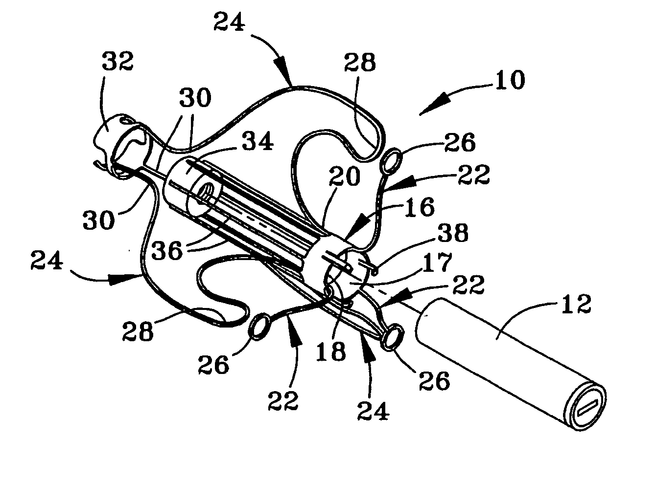
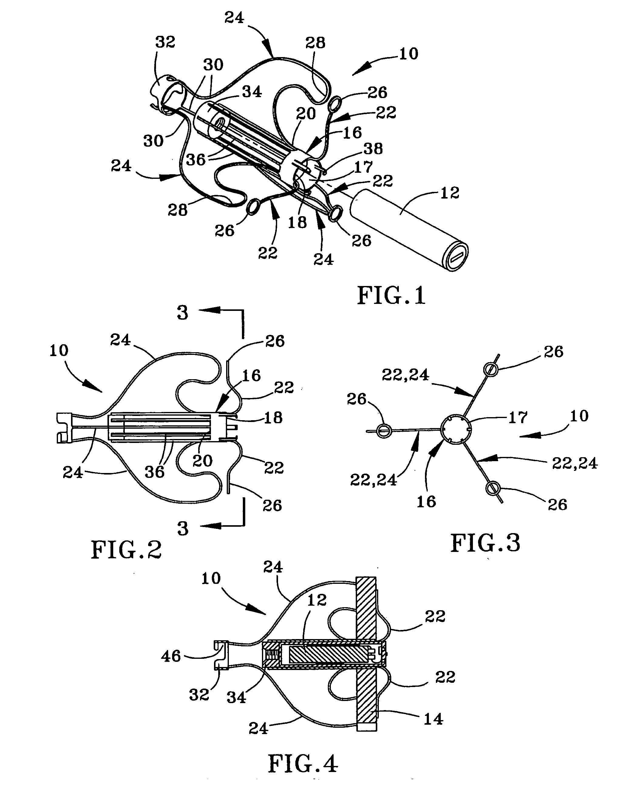
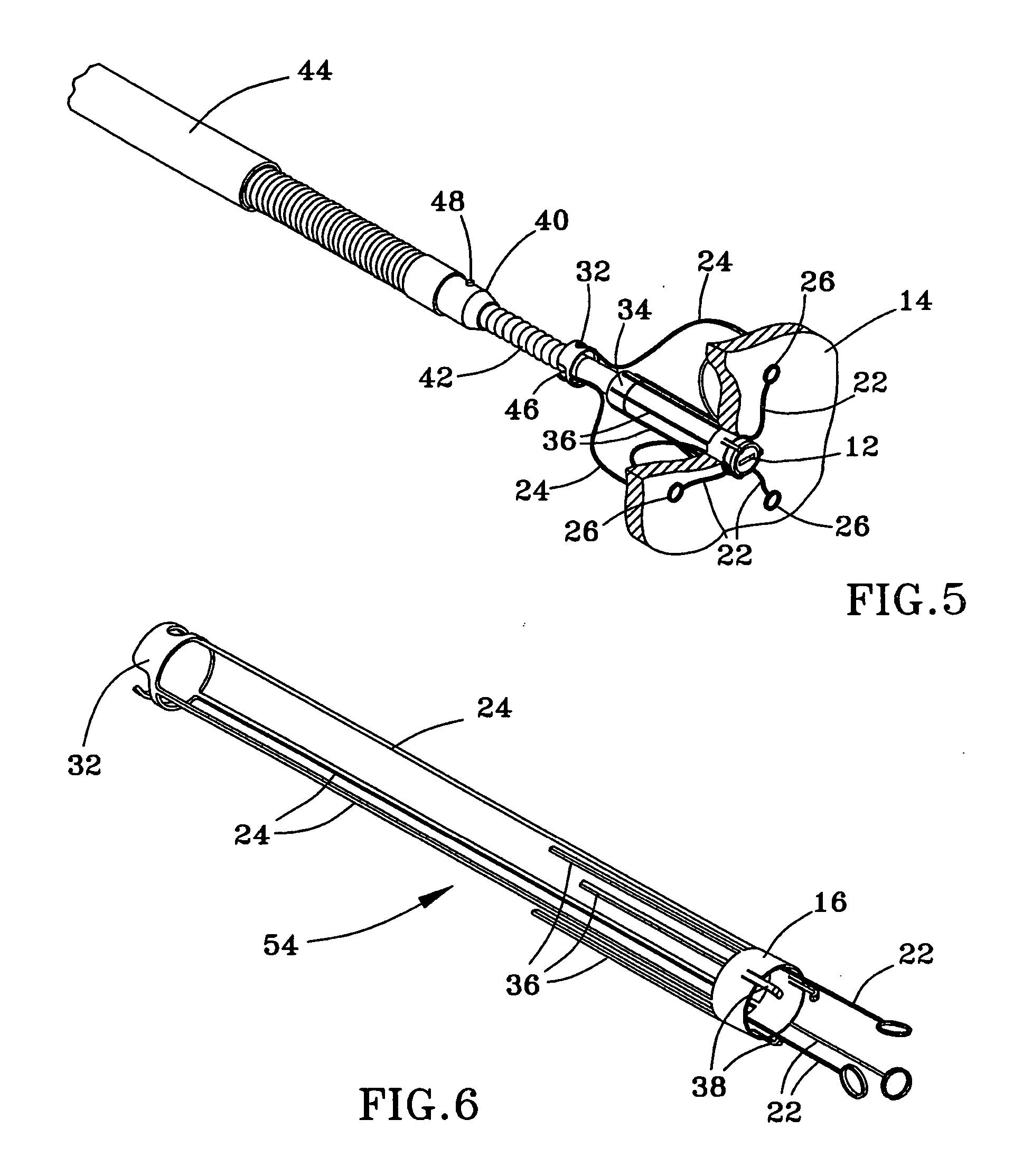
[0021]FIGS. 1 through 5 depict an anchor 10 suitable for delivering and securing a medical implant 12 to a wall 14, such as a wall of a cardiovascular organ, in accordance with an embodiment of the present invention. In a notable example, the wall 14 is an atrial septum and the implant 12 measures physiological parameters of the heart, such as LVEDP or MLA pressure. The implant 12 may be any one of a variety of types of implants currently known or developed in the future, and the scope of the present invention is not limited in any way by the type and operation of the implant 12.

[0022] The anchor 10 is represented in the Figures as having an annular-shaped central body 16 that defines a bore 17 in which the implant 12 is received. The central body 16 has oppositely-disposed first and second ends 18 and 20 corresponding to oppositely-disposed first and second directions along the axis of the central body 16. Arcuate arms 22 extend substantially radially and in the first direction fr...


PUM


Abstract

An anchor and procedure for placing a medical implant, such as for monitoring physiological parameters. The anchor includes a central body in which a medical implant can be received. arms and members extend radially from first and second ends, respectively, of the central body. Each member defines a leg extending toward distal portions of the arms to provide a clamping action. The anchor and its implant are placed by coupling first and second guidewires to first and second portions of the anchor, placing an end of a delivery catheter in a wall where implantation is desired, inserting the anchor in the catheter with the guidewires to locate the anchor within the wall, deploying the arms of the anchor at one side of the wall followed by deployment of the members at the opposite side of the wall, and thereafter decoupling the guidewires from the anchor.

Description

CROSS REFERENCE TO RELATED APPLICATIONS [0001] This application claims the benefit of U.S. Provisional Application No. 60 / 489,974, filed Jul. 25, 2003, and 60 / 491,002, filed Jul. 30, 2003.STATEMENT REGARDING FEDERALLY SPONSORED RESEARCH [0002] This invention was made with Government support under Contract No. NNC04CA10C awarded by National Astronautics and Space Agency. The Government has certain rights in the invention.BACKGROUND OF THE INVENTION [0003] (1) Field of the Invention [0004] The present invention relates generally to implantable medical devices, such as of the types for monitoring physiological parameters. More particularly, the invention relates to an anchor for a medical implant, a method of manufacturing anchors, and a method of placing a medical implant, wherein the anchor and method are suitable for use in procedures performed to diagnose, monitor, and / or treat cardiovascular diseases, including procedures performed to monitor pressure in the left side of the heart...

Claims


Application Information

Patent Timeline
Patent Type & Authority Applications(United States)
IPC IPC(8): A61B5/0215A61B17/00A61B17/12A61N1/05
CPCA61B5/0215A61B17/0057A61B17/12099A61B17/12122A61B17/12172A61B2017/00575A61B2017/12095A61B2017/00606A61B2017/00623A61B2562/028A61N1/057A61B2017/12054A61B2017/00592
Inventor SCHNEIDER, RICHARD LEENAJAFI, NADERGOETZINGER, DAVID J.
Owner UIM PRESSURE IMPLANT INC
Why Eureka
Try Eureka