Method and apparatus for co-display of inverse mode ultrasound images and histogram information

a technology of inverse mode ultrasound and histogram information, which is applied in the field of ultrasonic methods and apparatuses for analyzing a region of interest, can solve the problems of inability to combine images and certain types of non-image information in an easily viewable and adjustable manner

Inactive Publication Date: 2005-12-08
GE MEDICAL SYST GLOBAL TECH CO LLC
View PDF10 Cites 82 Cited by
  • Summary
  • Abstract
  • Description
  • Claims
  • Application Information

AI Technical Summary

Problems solved by technology

However, conventional systems have been unable to combine images and certain types of non-image information in an easily viewable and adjustable manner.

Method used


Image

  • Method and apparatus for co-display of inverse mode ultrasound images and histogram information
  • Method and apparatus for co-display of inverse mode ultrasound images and histogram information
  • Method and apparatus for co-display of inverse mode ultrasound images and histogram information

Examples


Embodiment Construction

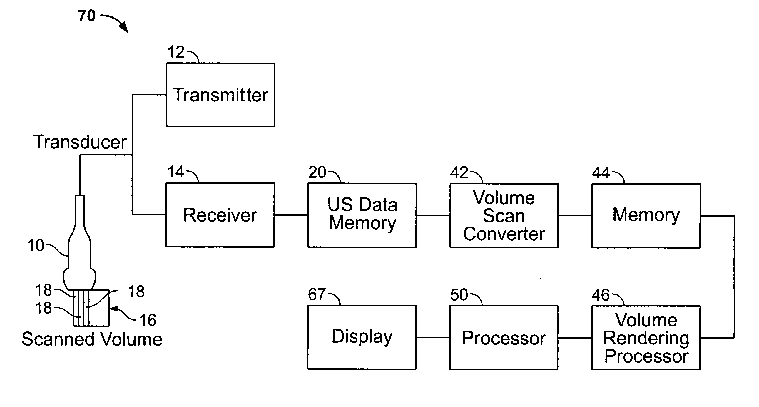
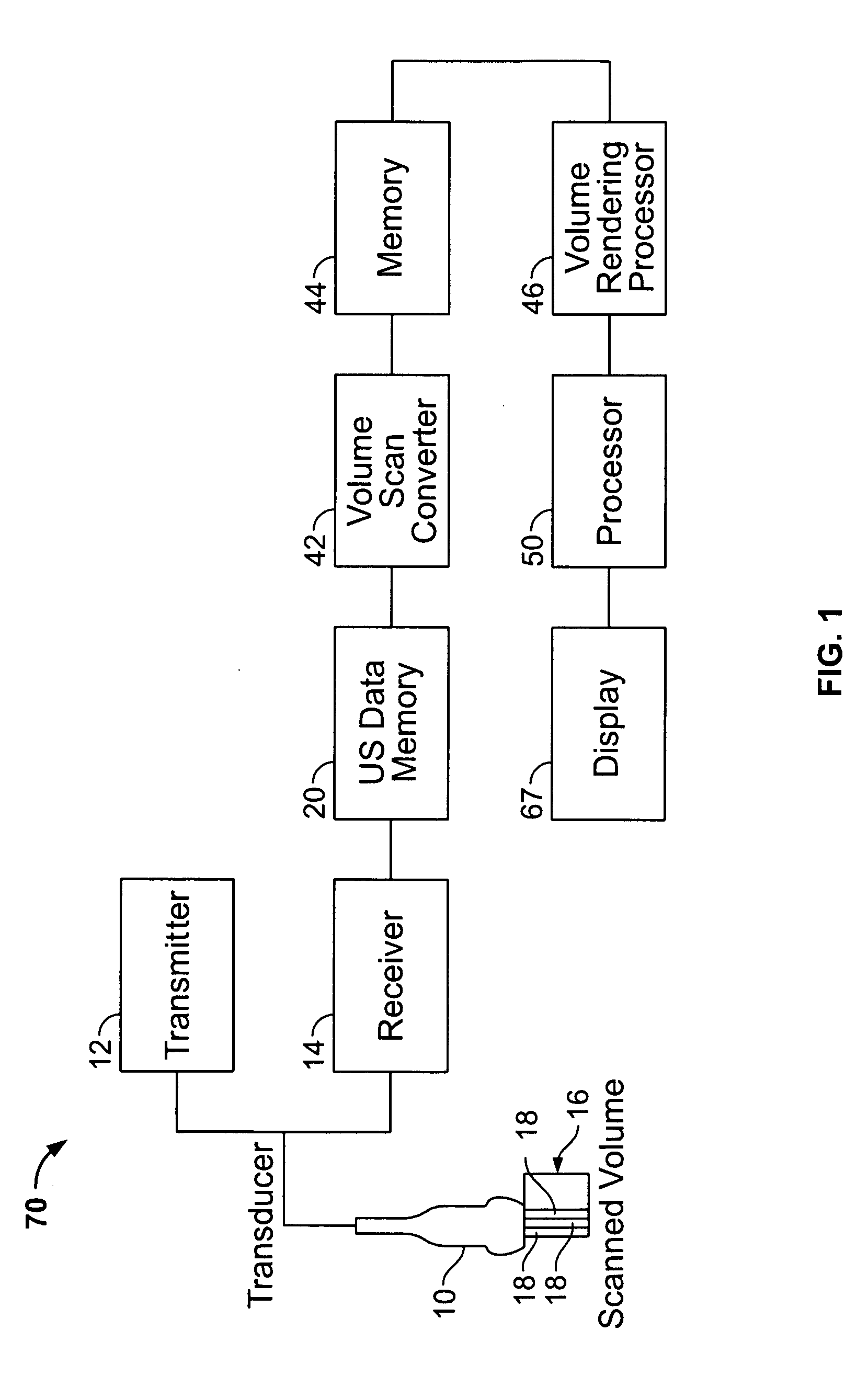
[0018]FIG. 1 illustrates an ultrasound system 70 formed in accordance with one embodiment of the present invention. The system 70 includes a probe 10 connected to a transmitter 12 and a receiver 14. The probe 10 transmits ultrasonic pulses and receives echoes from structures inside of a scanned ultrasound volume 16. Memory 20 stores ultrasound data from the receiver 14 derived from the scanned ultrasound volume 16. The volume 16 may be obtained by various techniques (e.g., 3D scanning, real-time 3D scanning, 2D scanning with transducers having positioning sensors, freehand scanning using a voxel correlation technique, 1.25D, 1.5D, 1.75D, 2D or matrix array transducers and the like).

[0019] The probe 10 is moved, such as along a linear or arcuate path, or electronically steered when using a 2D array, while scanning a region of interest (ROI). At each linear or arcuate position, the transducer 10 obtains scan planes 18. The scan planes 18 are stored in the memory 20, and then passed t...


PUM


Abstract

An ultrasound system is provided for analyzing a region of interest. The ultrasound system includes a probe for acquiring ultrasound information associated with the region of interest and a memory for storing a volumetric data set corresponding to at least a subset of the ultrasound information for at least a portion of the region of interest. The system further includes at least one processor for generating histogram information based on the volumetric data set and for generating ultrasound images based on the volumetric data set. The processor formats the histogram information and the ultrasound images to be co-displayed. The system further includes a display for simultaneously co-displaying the histogram information and the ultrasound images.

Description

BACKGROUND OF THE INVENTION [0001] The present invention generally related to an ultrasound method and apparatus for analyzing a region of interest and more particularly to a method and apparatus for co-displaying inverse mode ultrasound images and histogram information. [0002] Ultrasound systems have long existed for analyzing various regions of interest, such as in medical applications and in non-medical fields. Conventional ultrasound systems display the ultrasound information in a variety of formats and configurations. By way of example, existing ultrasound systems may display a series of two dimensional images or slices based on a volume of acquired data where the position of each slice is determined by the user. Along with the set of two dimensional slices or images, a rendered image (e.g. a three dimensional representation) may be separately or simultaneously displayed with one or more of the two dimensional images or slices. Conventional systems provide the user with various...

Claims


Application Information

Patent Timeline
Patent Type & Authority Applications(United States)
IPC IPC(8): G03B42/06A61B8/00
CPCA61B8/00A61B8/463A61B8/483
Inventor DEISCHINGER, HARALDBRANDL, HELMUT
Owner GE MEDICAL SYST GLOBAL TECH CO LLC
Why Eureka
Try Eureka