
System and method for measuring the rate of flow of cerebral spinal fluid into an external ventricular drainage mechanism

a technology of cerebral spinal fluid and rate of flow, which is applied in the field of system and method of measuring the rate of flow of cerebral spinal fluid into an external ventricular drainage mechanism, can solve the problems of patient's brain, csf that should flow from the brain to be trapped, injury or even death,

Inactive Publication Date: 2006-11-23
CALUORI RAYMOND J
View PDF4 Cites 3 Cited by
  • Summary
  • Abstract
  • Description
  • Claims
  • Application Information

AI Technical Summary

Benefits of technology

[0013] The alarm may be electrically powered or battery powered, or electrically powered with a battery back up. The battery back up will allow caregivers and patients to disconnect the measurement / alarm system from a wall socket and still operate on the battery for some period of time. This alarm device will allow the medical caregivers to know when the manual on / off valve may have been left in the off position by sounding after a predetermined time period.

Problems solved by technology

Cerebral shunts are placed in patients who have either an inability to absorb CSF, or when flow of CSF from the ventricles of the brain is obstructed, causing CSF that should flow from the brain to be trapped.
This increased pressure, if not released, can have many negative effects on the patient's brain that can cause injury or even death.
The position of the EVDM relative to the patients head is set avoid column pressure and column vacuum in the drainage tube: if the EVDM were positioned above the head, column pressures would increase, causing the need for more pressure for the CSF to leave the brain, which would create higher ICP, and possible patient discomfort or injury.
If the EVDM were positioned below the head, this could cause a lower vacuum pressure in the drainage tube, resulting in the removal of too much CSF from the brain, which can cause low ICP and possible patient discomfort or injury.
There are problems with the monitoring of CSF in the current usage of EVDMs.
One problem is there is no alarm.
This can lead to discomfort, brain injury and ultimately death by crushing the brain if not caught in time.
Another problem with EVDMs is how the volume and rate of flow of CSF from a patient's brain are determined.
This can be very hard to monitor accurately because typically the caregiver is a nurse who is caring for several patients at the same time, making it difficult for the nurse to be present to take measurements right on the hour.
This leads to inaccuracies in the monitoring and recording of the actual CSF flow rate and collection volume.

Method used


Image

  • System and method for measuring the rate of flow of cerebral spinal fluid into an external ventricular drainage mechanism
  • System and method for measuring the rate of flow of cerebral spinal fluid into an external ventricular drainage mechanism
  • System and method for measuring the rate of flow of cerebral spinal fluid into an external ventricular drainage mechanism

Examples


embodiment 40

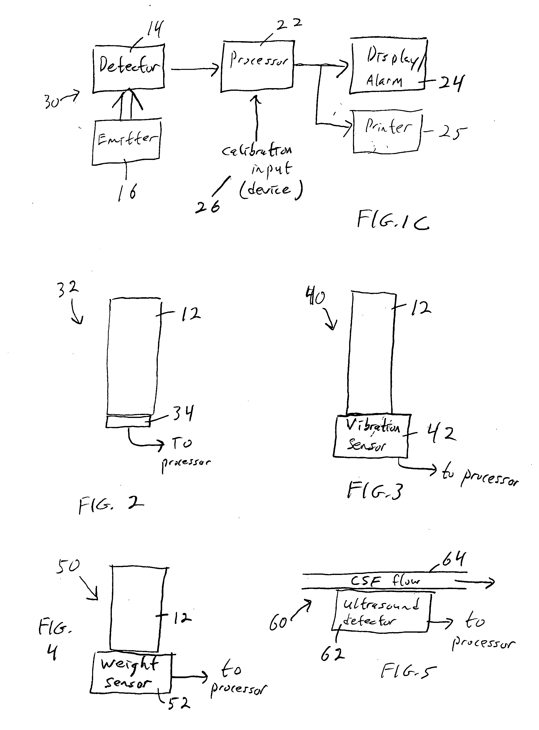
[0034] Yet another alternative embodiment 40, FIG. 3, is based on vibration. A drop of CSF falling into a collected volume in the EVDM collection apparatus (e.g. collection tube 12) causes a vibration in the volume. A sensor 42 that is excited with every drop that falls into the fluid (e.g. a pressure sensor) is connected to a microprocessor that counts the vibrations and calculates the rate and flow of CSF during a given period of time. If no vibrations are detected for a given period of time an alarm will activate.

[0035] This arrangement would employ a sensor that is on the inside of the tube, but with leads passing through the tube, the CSF will remain sterile. This method would allow the CSF to always be in a sterile environment and have no interaction with anything outside of the sterile interior of the EVDM.

embodiment 50

[0036] Yet another alternative embodiment 50, FIG. 4, works by detecting a change in mass of the collected CSF volume. Every time a drop of CSF falls into the collection tube 12, the total weight of the tube plus collected CSF increases. In other words, this arrangement would sense that the whole collection tube just gotten heavier. The weight is determined by sensor 52, which could be a scale or strain gauge, for example. A microprocessor then calculates the flow rate and total volume of CSF collected from the weight of the CSF that has been collected, and the weight change over time. If no weight change is detected for a given period of time, an alarm will be activated.

[0037] This arrangement would employ sensors that are on the exterior of the sterile EVDM collection container. This method would allow the CSF to always be in a sterile environment and have no interaction with anything outside of the sterile interior of the EVDM.

[0038] An alternative weight-based arrangement conte...

embodiment 70

[0041] Another embodiment 70, FIG. 6, uses changes in resistance. There are two ways to employ this arrangement. In a first method, every time the volume of a drop of CSF is added to the collected volume of CSF in tube 12, a slight change in the electrical conductivity, or resistance, of the total volume of fluid will be detected. Electrodes 72 and 74 are used for this purpose. With each additional volume (drop) of CSF added to the total volume collected, the conductivity will change. Again, a microprocessor can from these readings calculate the CSF flow rate and total collected volume. If there are no resistance changes sensed to the collected volume of CSF for a given period of time, an alarm will activate. This method may employ sensors and leads that are on the interior or exterior of the sterile EVDM's collection container. This method would allow the CSF to always be in a sterile environment and have no interaction with anything outside of the sterile interior of the EVDM.

[004...


PUM


Abstract

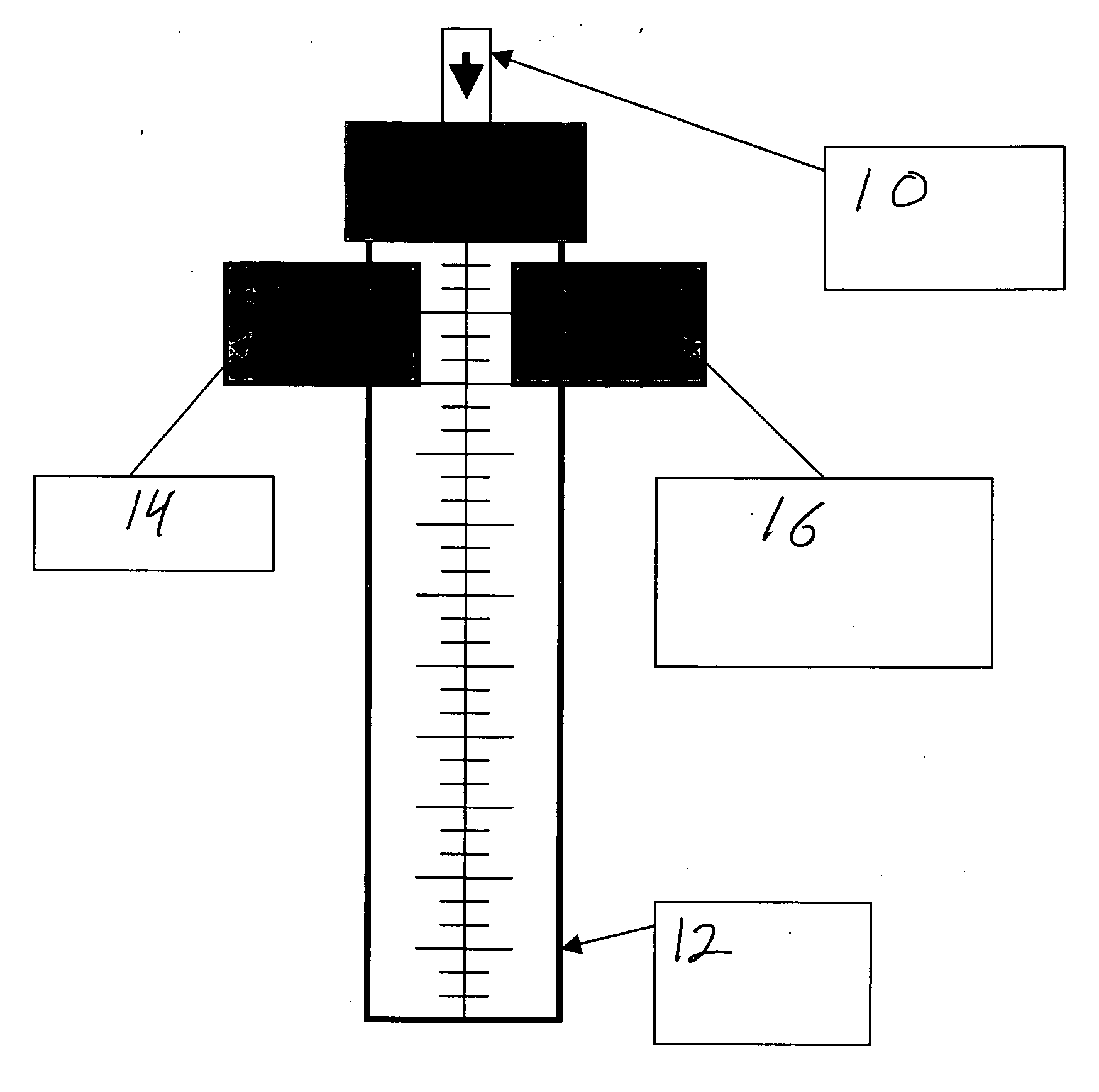
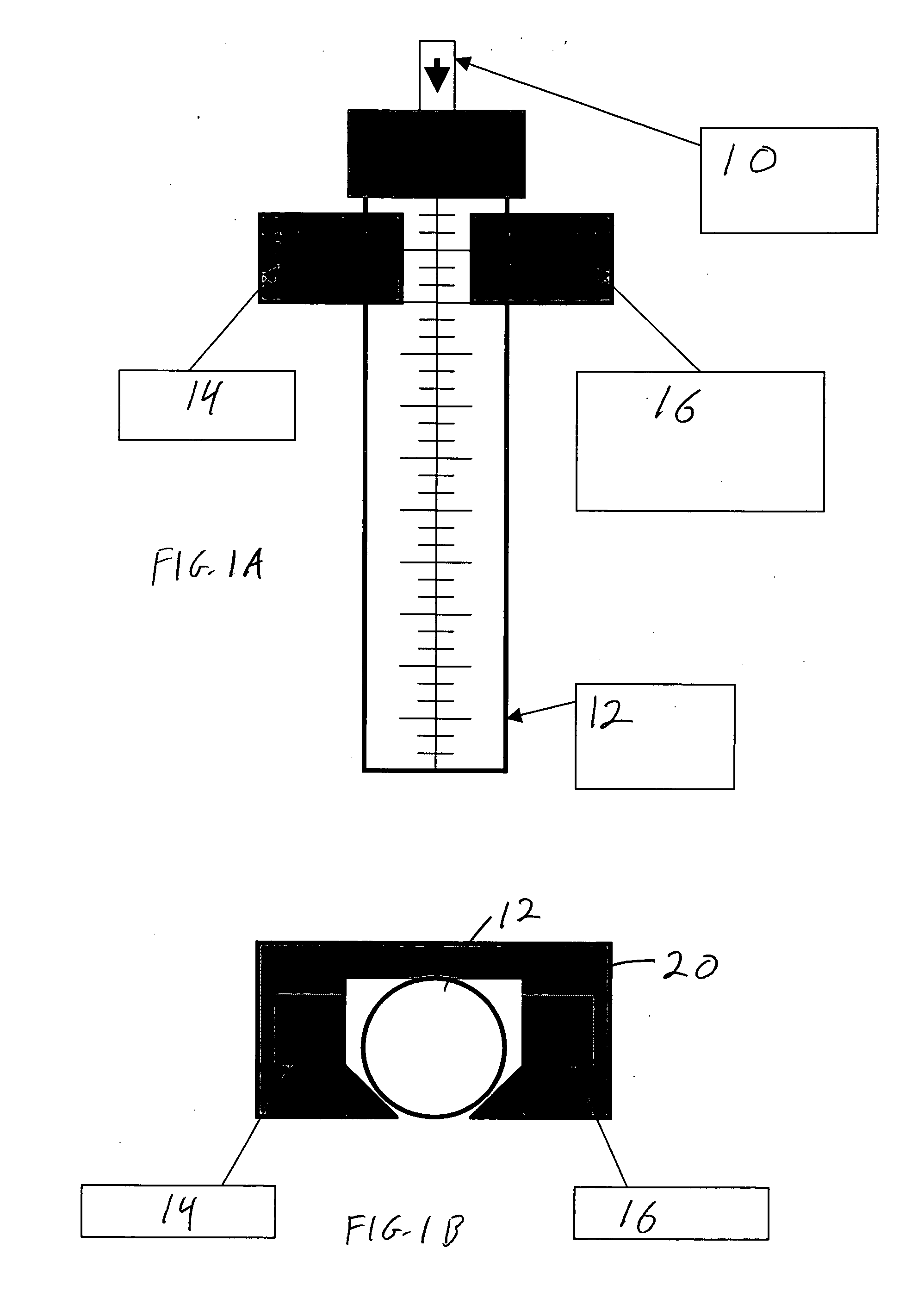
A system and method of determining the rate of flow of Cerebral Spinal Fluid (CSF) in an External Ventricular Drainage Mechanism (EVDM). Drops, or the flow of CSF moving within or into the EVDM, are detected. The user is provided, in a human-readable form, information derived from the detection. This may be accomplished with a printer and / or a digital display device. The information may include CSF flow rate and / or CSF volume collected in the EVDM, for example. The system and method may further determine if the drops or flow have ceased for at least a certain amount of time. In that case, an alarm may be activated.

Description

CROSS REFERENCE TO RELATED APPLICATION [0001] This application claims priority of Provisional application Ser. No. 60 / 683,674, filed on May 23, 2005.FIELD OF THE INVENTION [0002] This invention relates to a system and method of measuring the rate of flow of cerebral spinal fluid into an external ventricular drainage mechanism. BACKGROUND OF THE INVENTION [0003] A cerebral shunt works by regulating the pressure at which Cerebral Spinal Fluid (CSF) is allowed to leave the brain and taken to another part of the body for absorption. The brain produces approximately 18 ml / hour of CSF. Total CSF in circulation in an adult is approximately 140 ml, with 25 ml in the ventricles and 115 ml in the subarachnoid space. Cerebral shunts are placed in patients who have either an inability to absorb CSF, or when flow of CSF from the ventricles of the brain is obstructed, causing CSF that should flow from the brain to be trapped. Trapped fluid increases the Intra-Cranial Pressure (ICP). This increase...

Claims


Application Information

Patent Timeline
IPC IPC(8): A61M5/00
CPCA61B5/4261A61M27/006A61B5/4283
Inventor CALUORI, RAYMOND J.
Owner CALUORI RAYMOND J
Learn More