Interactive Radiological sciences clinical training system

a radiological science and clinical training technology, applied in the field of radiological science medical training devices, can solve the problems of increasing the cost of proper medical education and training, the cost of a qualified instructor or proctor is responsible for a significant component of these costs, and the training of practitioners is relatively small, so as to facilitate education and training.

Inactive Publication Date: 2006-12-14
MCCLAIN LOLITA A
View PDF7 Cites 11 Cited by
  • Summary
  • Abstract
  • Description
  • Claims
  • Application Information

AI Technical Summary

Benefits of technology

[0024] In accordance with yet another embodiment of the present invention, multiple video windows may be provided showing differing, yet simultaneous, views of a radiological science procedure. In this regard, a video window may be provided showing the external view of a radiological science procedure, while a second video window may illustrate the procedure as viewed internally through an optical lens. Yet a third window may be provided to graphically illustrate a cross-sectional side view of the tissue in the area where the medical procedure is to be performed. It will be appreciated that other similar video or visual windows may be provided on the display to facilitate the education and training consistent with the concepts of the present invention.

Problems solved by technology

In recent years, there has been an ever-growing increase in the expense of proper medical education and training.
The high cost of a qualified instructor or proctor is responsible for a significant component of these costs.
Relatively little investment has gone into the training of practitioners for the proper and safe performance of these procedures, notwithstanding the progress in the development of technology and equipment for advanced radiological science procedures.
While this method may provide a viable option for medical students, interns, or resident practitioners, it is generally not a feasible option for most licensed general surgeons, due to the time and expense involved.
Alternatively, a one or two day short course comprising lecture, video, and / or observation does not provide adequate training for more advanced medical procedures.
While some radiological science practitioners, particularly in urban areas, may be fortunate enough to establish proctorships with a radiological science instructor at a training center, these proctorships are often time prohibitive and fail to reach enough practitioners.
While the use of videotapes effectively reduces the cost associated with a live instructor or proctor, the limitations of video include the lack of interaction.
In short, training exclusive by way of video fails to permit the free exchange of question and answer, and avoids the “hands-on” training that is essential to any quality educational / training program.
Although the use of mannequins provides an effective method of achieving the coordinative skills of some radiologic science procedures, the anatomical divergence between a live human and a mannequin generally dampens the learning curve.
Moreover, the use of mannequins is often very expensive, since universities, colleges, and other training institutions may be able to afford the actual mannequin but, not the multi million dollar radiological science equipment to perform the procedures itself.
These traditional methods of training require constant supervision by highly skilled and licensed technologist, thus, limiting the number of persons who may be competently trained to perform such new procedures in a specified amount of time.
Organizations which have sought to train medical personnel in developing countries have found these traditional methods of training especially frustrating because of the limited availability of skilled persons and necessary facilities to provide such training.
The number of persons which the organization may train effectively using these training techniques is limited to a few select radiological science student.
Consequently, the organization has not been able to adequately train a sufficient numbers of technologist to diagnose and treat such conditions as cancer, which currently affect an estimated thirty-five million people worldwide.

Method used


Image

  • Interactive Radiological sciences clinical training system
  • Interactive Radiological sciences clinical training system
  • Interactive Radiological sciences clinical training system

Examples


Embodiment Construction

[0031] By way of example, the present invention is illustrated in terms of a system for use in training medical personnel in radiological science procedures. The example application described herein is only one example application of the present invention and is provided for the purpose of better explaining the present invention. The present invention may be applied to any number of other radiological science procedures. Thus, the present invention should not be limited to the specific example of radiological science procedures described herein.

The Hardware

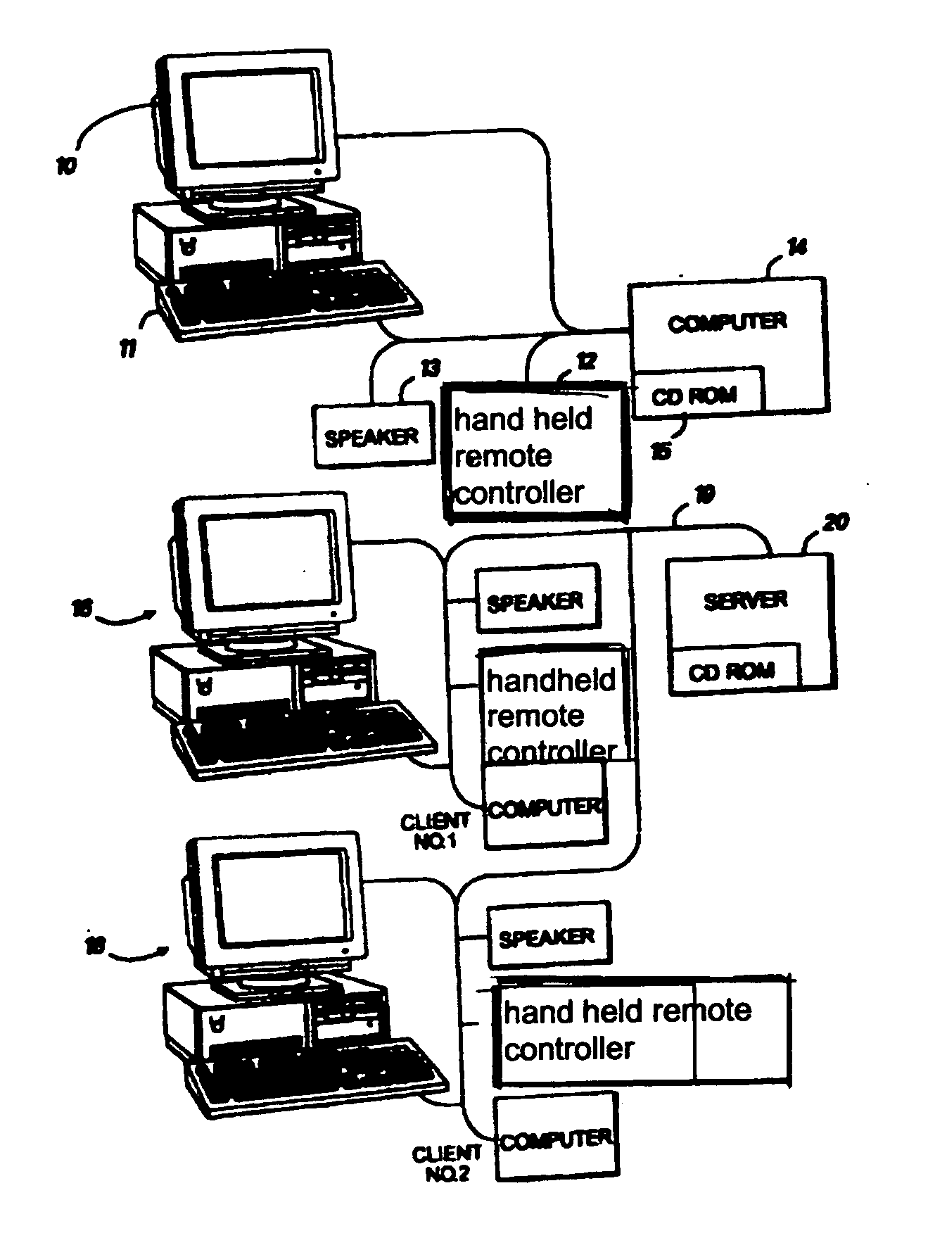
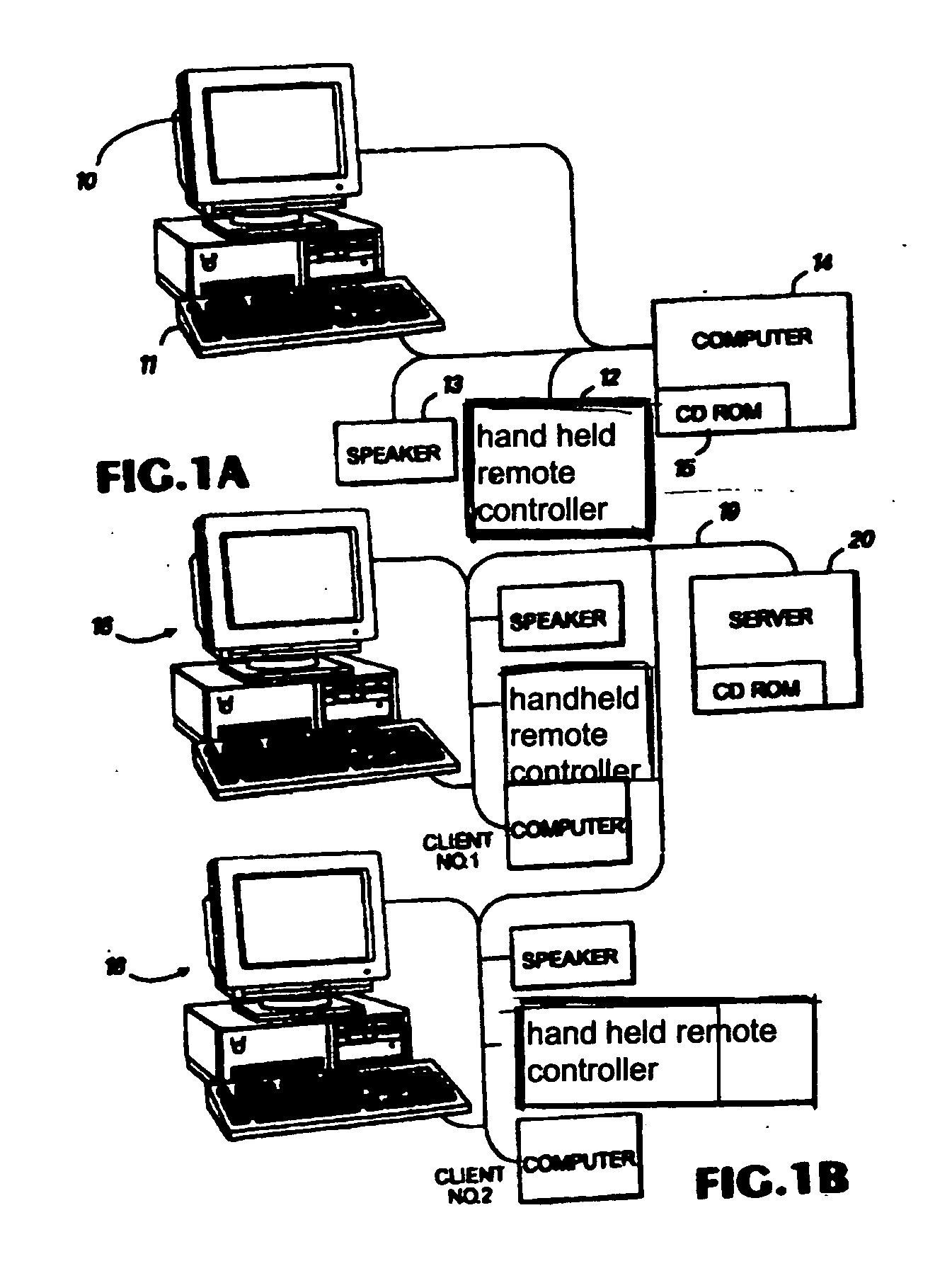
[0032] Shown in FIG. 1 is an example of a multimedia computer system 10 which could be employed to implement the present invention. In FIG. 1, a computer system 10 is shown having at least a 486 based 33 Mhz CPU based computer 1 with an internal hard drive, a disk drive 9 (which could also include a CD-ROM reader 8), a monitor 2, speakers 3 and a laser disk player 4 for playing a laser disk 5. The computer system 10 may also in...


PUM


Abstract

An interactive medical training device comprising a computer system having a display, wherein the computer system is programmed to provide education and training in radiological sciences, more particularly in radiological procedures that promote the diagnoses of many ailments and the treatment of cancer and/or tumors. The computer system simulates radiological procedures for medical practitioners, students, and patients. This aspect is achieved by configuring the system to display, on a portion of the display, a video window. The video window displays an interactive video segment illustrating a portion of the radiologic/radiation therapy procedure. The system requests a user to input information relating to a next step in the radiologic/radiation therapy procedure, which advantageously keeps the user engaged in the training session. This “next step” information may include, for example, selecting an appropriate medical instrument or selecting a location on the body for radiation therapy/radiologic procedure. The user inputs the requested information through an input device, such as a mouse, a keyboard, a touch-sensitive screen, or other input device, such as a remote control-like device. The system then receives and interprets the user input and informs the user as to whether the input is correct. Preferable, if the input is correct the system will display a prerecorded interactive video segment illustrating the next step of the radiologic/radiation therapy procedure.

Description

BACKGROUND OF THE INVENTION [0001] 1. Field of the Invention [0002] The present invention generally relates to radiological science medical training devices, and more particularly to a system that provides interactive medical training for diagnosing and treatment procedures. [0003] 2. Discussion of the Related Art [0004] In recent years, there has been an ever-growing increase in the expense of proper medical education and training. This recent trend spans the entire spectrum of medical disciplines, from podiatry to neurology. The high cost of a qualified instructor or proctor is responsible for a significant component of these costs. Minimizing, or at least reducing this cost, will reduce the overall cost of medical education and training. Therefore, various devices have been employed to reduce this “live” instructional cost component. To better illustrate the problem and devices employed to alleviate the problem, more specific reference will be made to the medical field involving ...

Claims


Application Information

Patent Timeline
Patent Type & Authority Applications(United States)
IPC IPC(8): G09B23/28
CPCG09B23/28G09B19/00
Inventor MCCLAIN, LOLITA A.
Owner MCCLAIN LOLITA A
Why Eureka
Try Eureka