
Radiopaque expandable body and methods

Inactive Publication Date: 2007-01-11
KYPHON
View PDF97 Cites 176 Cited by
  • Summary
  • Abstract
  • Description
  • Claims
  • Application Information

AI Technical Summary

Benefits of technology

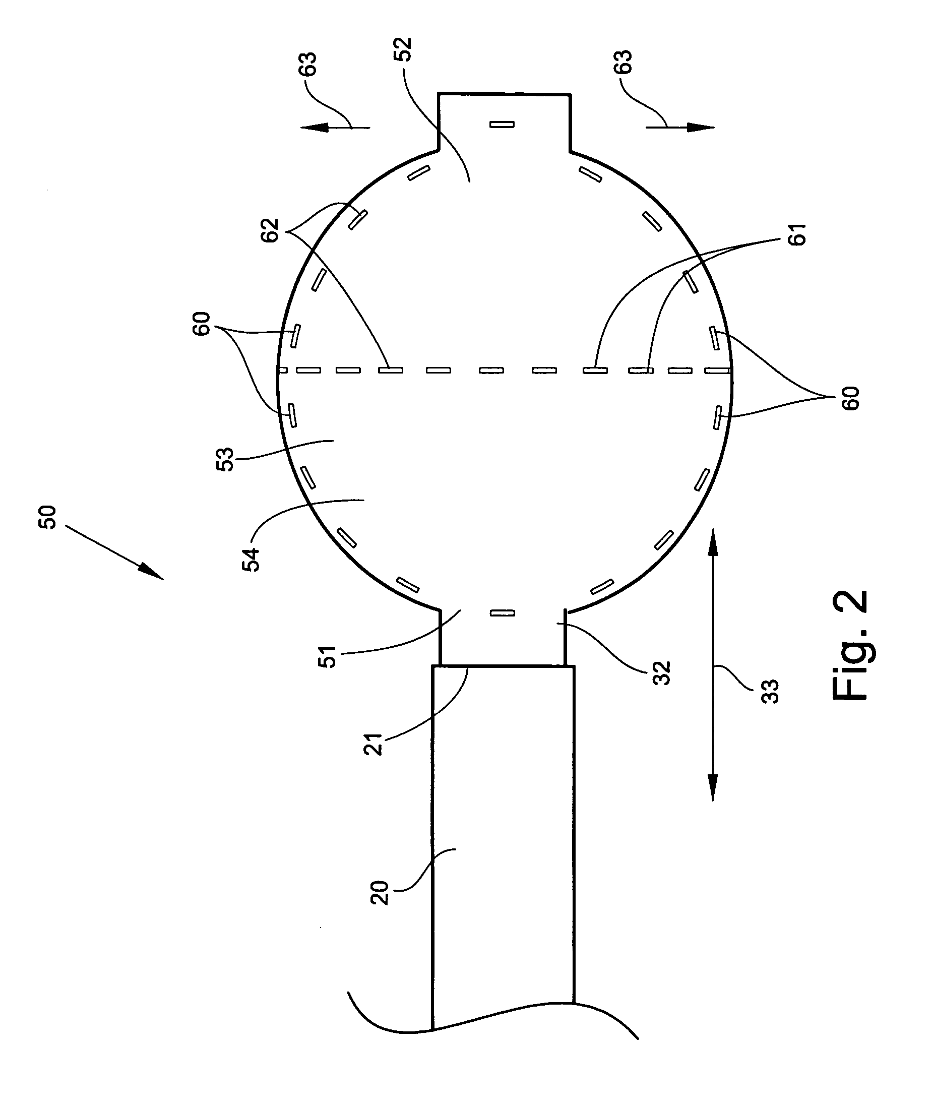
[0009] In an illustrative embodiment, the radiopaque marking pattern can include a plurality of radiopaque markers. Each of the radiopaque markers can be in communication with a predetermined location on the expandable body. As a result, the positioning—for example, the orientation and movement—of the expandable body in various directions can be visualized radioscopically. Such a device is useful for diagnostic or therapeutic purposes, including, for example, providing cavities in interior body regions.
[0010] Features of a radiopa

Problems solved by technology

These conditions, if not successfully treated, can result in deformities, chronic complications, and an overall adverse impact upon the quality of life.
A disadvantage of using radiopaque contrast media is that accidental exposure to such radiopaque media can cause hypersensitivity reactions in some patients.
Other disadvantages include difficulty in handling radiopaque media that are more viscous than, for example, a saline solution, and that radiopaque media are generally more expensive than non-radiopaque fluids.
However, the availability of MRI monitoring may be limited, and the use of MRI can be very expensive.

Method used


Image

  • Radiopaque expandable body and methods
  • Radiopaque expandable body and methods
  • Radiopaque expandable body and methods

Examples


Embodiment Construction

[0021] Embodiments of the present invention provide systems and methods for radioscopic visualization of the positioning of an expandable body in an interior body region. The systems and methods embodying the invention can be adapted for use in many suitable interior body regions, wherever the formation of a cavity within or adjacent one or more layers of tissue may be required for a therapeutic or diagnostic purpose. The illustrative embodiments show the invention in association with systems and methods used to treat bones. In other embodiments, the present invention may be used in other interior body regions or types of tissues.

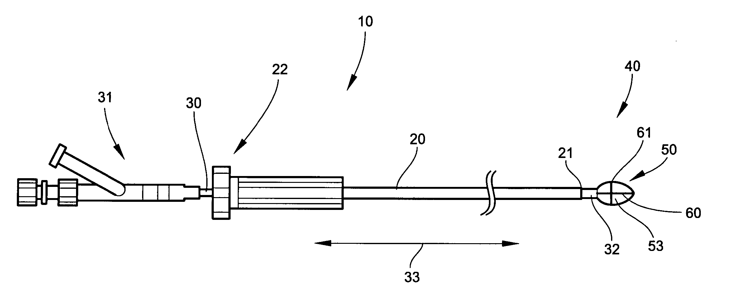
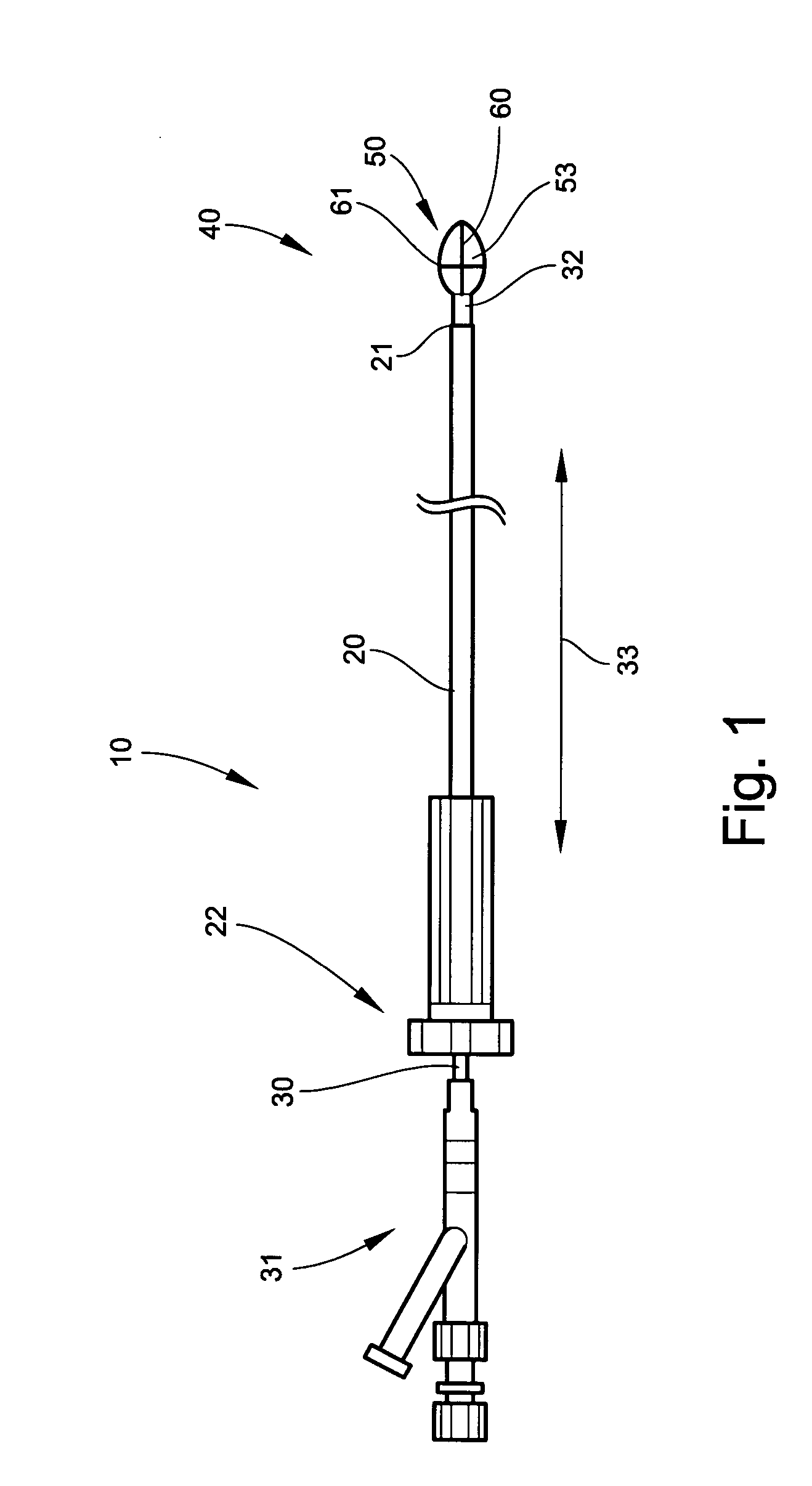
[0022] Referring now to the figures, FIG. 1 is a view of a system 10 according to an embodiment of the present invention configured to allow an user to provide a cavity in a targeted treatment area in an interior body region. The system 10 includes an expandable body 50 configured to be used in a kyphoplasty procedure. Kyphoplasty is a minimally invasive s...


PUM


Abstract

Systems and methods for radioscopic visualization of the positioning of an expandable body in an interior body region are described. One such system includes a radiopaque marking pattern in communication with the expandable body. The radiopaque marking pattern can be configured to allow for visualizing radioscopically the orientation and movement of the expandable body in unexpanded and expanded states in an interior body region. Such a device is useful for diagnostic or therapeutic purposes, including, for example, providing cavities in interior body regions.

Description

FIELD OF THE INVENTION [0001] The present invention relates to systems and methods for radioscopic visualization of the positioning of an expandable body in an interior body region. Such systems and methods may be useful for diagnostic or therapeutic purposes, for example, creating a cavity in a vertebral body. BACKGROUND OF THE INVENTION [0002] Certain diagnostic or therapeutic procedures require provision of a cavity in an interior body region. For example, as disclosed in U.S. Pat. Nos. 4,969,888 and 5,108,404, a balloon may be deployed to form a cavity in cancellous bone tissue as part of a therapeutic procedure that fixes fractures or other abnormal bone conditions. The balloon or other expandable body may compress the cancellous bone to form an interior cavity. A filling material, such as a bone cement, may be inserted into the cavity in order to provide interior structural support for cortical bone. [0003] This procedure can be used to treat cortical bone, which—due to osteop...

Claims


Application Information

Patent Timeline
IPC IPC(8): A61M29/00
CPCA61B17/8855A61M2025/1079A61M25/10A61M25/0108
Inventor GONG, GORMANOSORIO, REYNALDO A.
Owner KYPHON
Learn More