Method for generating an image exposure of the heart requiring a preparation

a technology of image exposure and preparation, applied in the field of creating an image exposure of the heart of an examination, can solve the problems of repeat acquisition, limited image quality, and possible inability to use acquired measurement,

Inactive Publication Date: 2007-03-29
SIEMENS AG
View PDF8 Cites 5 Cited by
  • Summary
  • Abstract
  • Description
  • Claims
  • Application Information

AI Technical Summary

Benefits of technology

[0027] Preparation pulses can be applied in the framework of the preparation. For example, given the image acquisition with a magnetic resonance apparatus radio-frequency pulses effect a saturation of the blood such that the signal of the blood is suppressed in the image exposures. The measurement is thereby prepared, for example in the dark blood technique. Other preparation methods can alternatively or additionally be used via which the slice or heart position relevant for the image acquisitions is likewise provided.

Problems solved by technology

Such errors lead to limitations of the image quality, such that the acquired measurement possibly may not be usable and a repeat acquisition is required.
The idle period in an examination apparatus (such as a scanner in magnetic resonance tomography) is extended and the system thus is blocked longer, with the result that subsequent examinations can be delayed.
Errors due to an incorrect manual parameterization also cannot be completely precluded even when the operator (such as the technical assistants or doctors) I well-trained, such that the image quality is subject to significant fluctuations dependent on the person who happens to be the operator and anomalies in the patient and measurements must frequently be repeated.

Method used


Image

  • Method for generating an image exposure of the heart requiring a preparation
  • Method for generating an image exposure of the heart requiring a preparation
  • Method for generating an image exposure of the heart requiring a preparation

Examples


Embodiment Construction

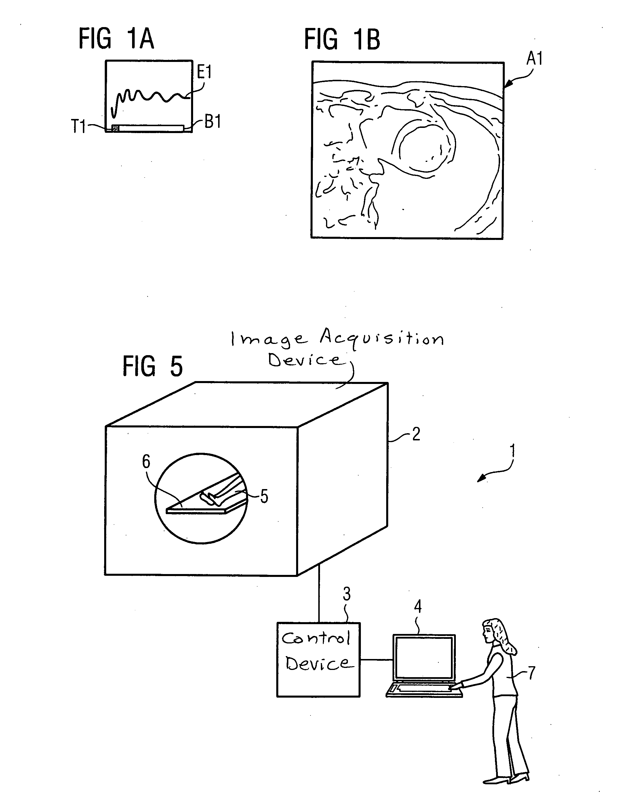
[0037]FIG. 1A shows an electrocardiogram E1 with a time bar B1. In the time bar B1 a point in time T1 is marked relatively early in the representation, at which point in time T1 an image acquisition is established directly following a preparation. An image data acquisition at the marked point in time T1 leads to the heart image of the image exposure A1 shown in FIG. 1B, from which the heart position relevant for the actual image acquisition is determined. The actual image acquisition with the imaging medical examination apparatus is to be implemented later at this heart position of FIG. 1B.

[0038] In the further course of the heart cycle the current heart position is determined from time to time via the electrocardiogram with the aid of the controller.

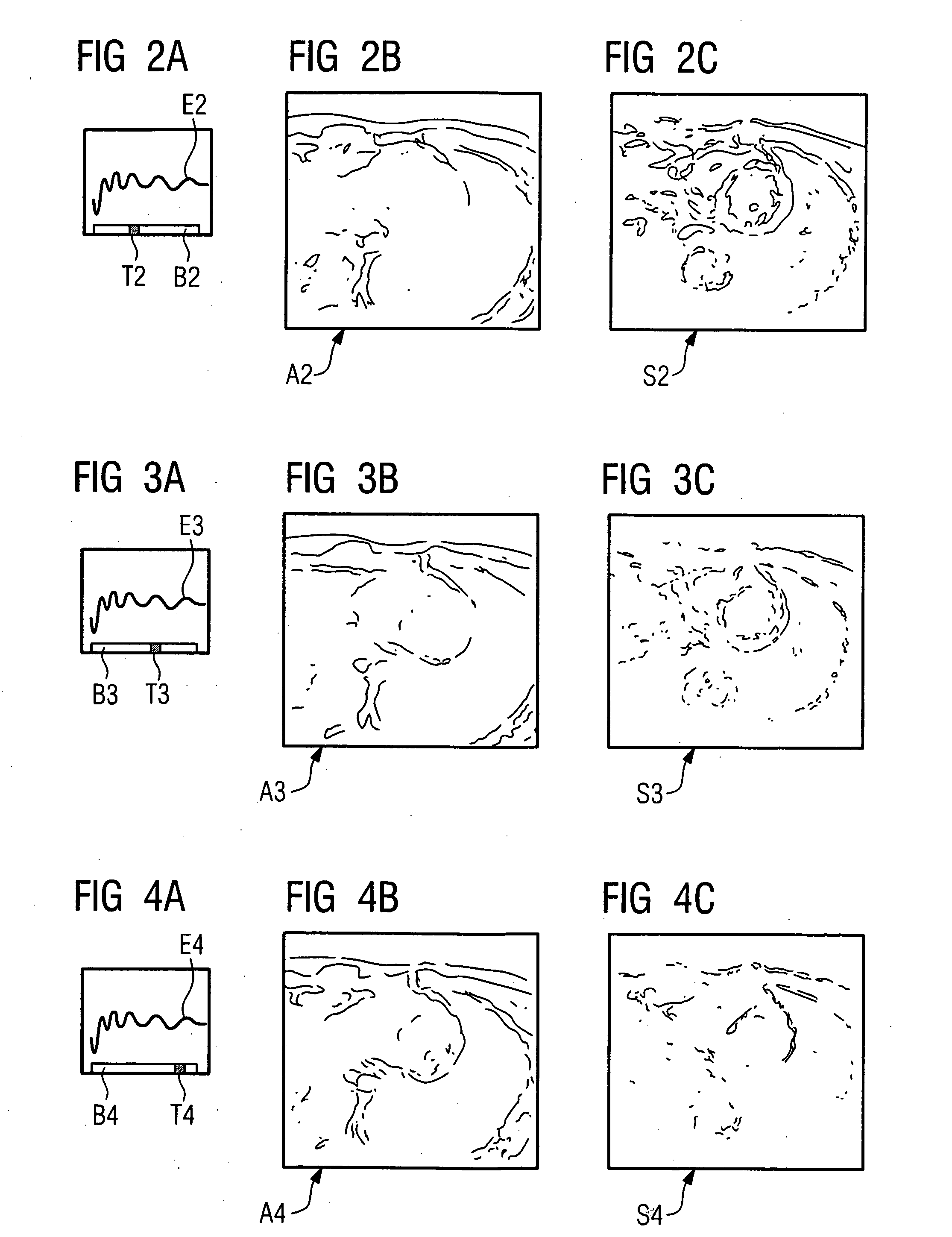
[0039] In FIG. 2A an electrocardiogram E2 is shown below which a time bar B2 is shown in which a point in time T2 is marked that, in comparison to the point in time T1 of the time bar B1, is to be associated with a later point in time...


PUM


Abstract

A method for generation of an image exposure of the heart of an examination subject with an imaging medical examination apparatus in particular a magnetic resonance apparatus, the image exposure requiring preparation includes the steps of: preparation of the heart in a heart position that is relevant for the image acquisition and determination of the associated heart position, determination of at least one current heart position in the further course of the heart cycle, comparison of the determined current heart position with the heart position relevant for the image acquisition, and given correlation of the current heart position and the heart position relevant for the image acquisition, starting the image acquisition through a control device of the imaging medical examination apparatus.

Description

BACKGROUND OF THE INVENTION [0001] 1. Field of the Invention [0002] The present invention concerns a method for creating an image exposure of the heart of an examination subject with an imaging medical examination apparatus, in particular a magnetic resonance apparatus, wherein the image exposure requires a preparation. [0003] 2. Description of the Prior Art [0004] Often a preparation is necessary in order to generate heart image exposures should of a human or animal examination subject. For example, for what is known the “dark blood” technique, the signal of blood is suppressed and the surrounding tissue can be better detected, such that the heart muscle is better visible in the image representations. For this purpose, for example, preparation pulses for preparation of the actual measurement occurring in the end diastole are applied directly after the R-spike, which the highest positive amplitude of the electrocardiogram. [0005] If the image data acquisition should occur in the end...

Claims


Application Information

Patent Timeline
Patent Type & Authority Applications(United States)
IPC IPC(8): A61B5/05
CPCA61B5/0456A61B5/7285A61B5/055A61B5/352
Inventor SPRUNG, KATRIN CHRISTEL
Owner SIEMENS AG
Why Eureka
Try Eureka