Method and system for using computed tomography to test pulmonary function

a computed tomography and pulmonary function technology, applied in the field of methods and systems of determining pulmonary function, can solve the problems of significantly poorer spectral resolution than that of ct, more expensive and time-consuming than computed axial

Active Publication Date: 2007-04-19
VIRGINIA COMMONWEALTH UNIV
View PDF11 Cites 12 Cited by
  • Summary
  • Abstract
  • Description
  • Claims
  • Application Information

AI Technical Summary

Benefits of technology

[0006] This invention is brings together two emerging technologies, thoracic four dimensional computed tomography (4DCT) and deformable-image registration algorithms, to devise a new clinical test for lung functi

Problems solved by technology

Currently, pulmonary ventilation is performed using single photon emission computed tomography (SPECT), a nuclear medicine imaging procedure, which is more

Method used


Image

  • Method and system for using computed tomography to test pulmonary function
  • Method and system for using computed tomography to test pulmonary function

Examples


Embodiment Construction

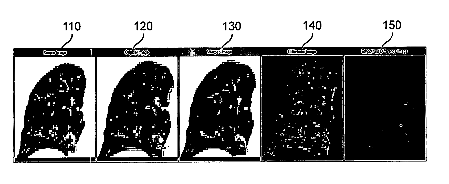
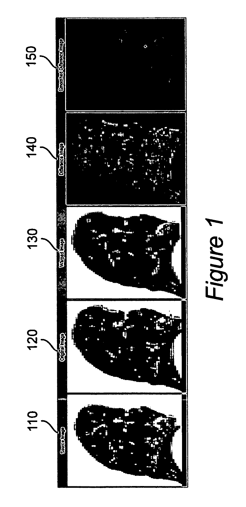
[0013] Four-dimensional computed tomography (4DCT) acquisition methods that explicitly account for respiratory motion have been recently developed in academic and commercial settings. Similarly, deformable image registration algorithms are evolving to the point of routine clinical utility. The combination of these two emerging technologies can be used as a pulmonary function test for ventilation by assessing the density differences of the same anatomic areas of the lung from CT scans acquired at different respiratory phases.

[0014] Due to the deformation of the lungs caused by respiration, the CT scans at different respiratory phases (referred to subsequently as inhale and exhale scans) cannot be directly compared as the anatomy is in a different location in the two images. FIG. 1a and 1b, respectively, show exhale 110 and inhale 120 images. However, deformable image registration algorithms can be applied to deform one respiratory phase to another. This is shown by the image 130 in ...


PUM


Abstract

Computed axial tomography images of different respiratory phases of lungs are obtained, where the intensity of the image measures lung density. One image is deformed to the coordinate space of the other image, and the differences between the intensity values of the other image as compared to the mapped image are evaluated as measures of ventilation.

Description

BACKGROUND OF THE INVENTION [0001] 1. Field of the Invention [0002] The present invention generally relates to methods and systems of determining pulmonary function, and in particular to methods and systems using computed tomography. [0003] 2. Background Description [0004] Currently, pulmonary ventilation is performed using single photon emission computed tomography (SPECT), a nuclear medicine imaging procedure, which is more expensive and time consuming than computed axial tomography (CAT scan, or CT). Furthermore, the resolution of SPECT is significantly poorer than that of CT. What is needed is a method for measuring lung ventilation that uses CT. SUMMARY OF THE INVENTION [0005] It is therefore an object of the present invention to measure pulmonary function using computed axial tomography (CT). [0006] This invention is brings together two emerging technologies, thoracic four dimensional computed tomography (4DCT) and deformable-image registration algorithms, to devise a new clin...

Claims


Application Information

Patent Timeline
IPC IPC(8): G06K9/00
CPCA61B6/50A61B6/5217G06T7/60G16H50/30
Inventor KEALL, PAUL JOHNJOSHI, SARANG C.
Owner VIRGINIA COMMONWEALTH UNIV
Why Eureka
Try Eureka