Medical image processing apparatus and medical image processing method

A processing device and medical image technology, applied in image data processing, image enhancement, image generation, etc., can solve problems such as judging the degree of COPD

Active Publication Date: 2015-11-25
CANON MEDICAL SYST COPRPORATION +1
View PDF11 Cites 13 Cited by
  • Summary
  • Abstract
  • Description
  • Claims
  • Application Information

AI Technical Summary

Problems solved by technology

[0005] However, it is difficult to accurately determine the degree of COPD only by using the above-mentioned ch

Method used


Image

  • Medical image processing apparatus and medical image processing method
  • Medical image processing apparatus and medical image processing method
  • Medical image processing apparatus and medical image processing method

Examples


Embodiment Construction

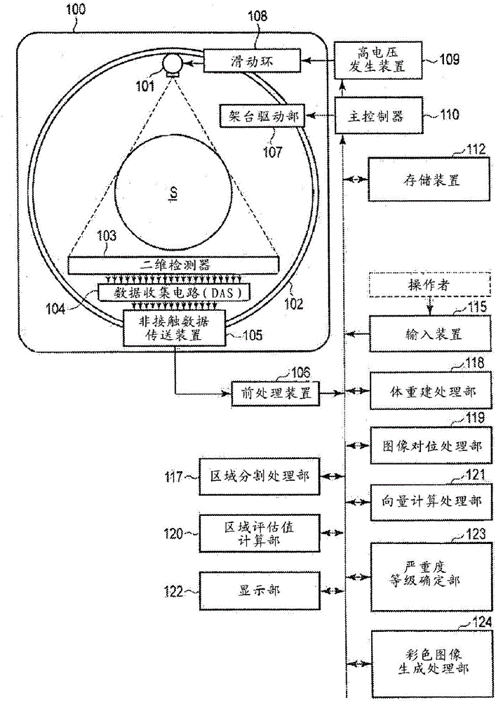
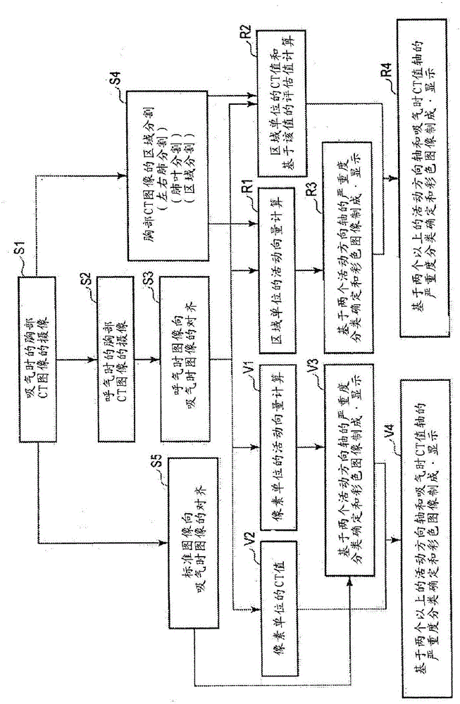
[0033] The medical image processing apparatus according to the present embodiment processes data of a plurality of images representing the shape of the subject's chest and having different respiratory phases. The amount of movement of a region between multiple images is calculated for each pixel or region. A level related to the severity of chronic obstructive pulmonary disease is determined for each pixel or region based on a combination of at least two of the activity amount, the feature value obtained from the pixel value of the image, and the rate of change in the size of the region. Information related to the determined level is output.

[0034] Hereinafter, the medical image processing apparatus according to the present embodiment will be described with reference to the drawings. In the following description, volume data representing the three-dimensional form (three-dimensional structure) of the inside of the chest of the subject is exemplified. This volume data is pr...


PUM


Abstract

The present invention provides information that is an effective aid in diagnosis of chronic obstructive pulmonary disease from thoracic volume images. The storage unit (112) of the medical image processing apparatus stores the data for multiple images of differing respiratory phases that represent the configuration of a subject's thoracic region. A vector-calculating unit (121) calculates the amount of movement of an area among the multiple images for each pixel or region. By combining at least two from among the amount of movement, a feature value determined from image pixel values and the rate of change in the size of a region, a level-determining unit (123) determines a level, which relates to the severity of chronic obstructive pulmonary disease, for each pixel or region.

Description

technical field [0001] Embodiments of the present invention relate to a medical image processing apparatus and a medical image processing method. Background technique [0002] The present invention relates to diagnosis of chronic obstructive pulmonary disease (COPD: Chronic Obstructive Pulmonary Disease), and particularly to an image diagnosis apparatus using chest CT images. COPD is a disease that includes chronic bronchitis and emphysema. In particular, it is the 4th leading cause of death in the United States after 3 major diseases (cancer, heart disease, and stroke) in the United States between the ages of 65 and 84, and is expected to surpass stroke by 2020. become the 3rd leading cause of death. Chronic bronchitis refers to the chronic inflammation of the trachea or the bronchi, the excessive secretion of mucus in the bronchi continuously or repeatedly, and the lumen of the bronchial structure is narrowed by fibrous tissue. In addition, emphysema is a state in which ...

Claims


Application Information

Patent Timeline
IPC IPC(8): A61B6/03A61B5/055
CPCA61B6/5264A61B5/055A61B6/5217A61B6/486A61B6/5229A61B6/5235G06T7/215A61B6/50G16H50/30A61B6/032G06T7/0012G06T11/003G06T15/08G06T2200/04G06T2207/10081G06T2207/30061G06T2210/41G06T2211/40
Inventor 山形仁大野良治
Owner CANON MEDICAL SYST COPRPORATION
Why Eureka
Try Eureka