Unlock instant, AI-driven research and patent intelligence for your innovation.

Method and device for measuring pulmonary nodule volume using dual-energy substraction image

a technology of pulmonary nodules and subtraction images, which is applied in the field of dual-energy subtraction images for measuring can solve the problems of difficult to accurately measure the volume of pulmonary nodules, difficult for medical experts to determine whether a pulmonary nodule is benign, and the volume measurement error is still about 10%, so as to achieve accurate measurement of pulmonary nodules.

Inactive Publication Date: 2007-06-14
ELECTRONICS & TELECOMM RES INST
View PDF7 Cites 1 Cited by
  • Summary
  • Abstract
  • Description
  • Claims
  • Application Information

AI Technical Summary

Benefits of technology

[0009] The present invention provides a method of accurately measuring a pulmonary nodule volume using a dual-energy subtraction image.
[0010] The present invention also provides a device for accurately measuring a pulmonary nodule volume using a dual-energy subtraction image.
[0012] In the above aspect of the present invention, (a) includes: (a1) obtaining high and low energy chest x-ray images; and (a2) removing x-ray absorption effect of bone tissues from the obtained images.
[0019] In the above aspect of the present invention, the dual-energy subtraction image obtaining unit: obtains high and low energy chest x-ray images; and removes an x-ray absorption effect of bone tissues from the obtained images.

Problems solved by technology

However, it is difficult even for medical experts to determine whether a pulmonary nodule is benign or malignant.
However, when the shape of the pulmonary nodule is not a sphere, it becomes difficult to accurately measure the volume of the pulmonary nodule.
When using a CT radiograph, the accuracy of measuring the pulmonary nodule volume is relatively high, but the volume measurement error is still about 10%.
When CT radiography is used, the volume measurement error is mainly caused by imperfect extraction of the pulmonary nodule border.

Method used


Image

  • Method and device for measuring pulmonary nodule volume using dual-energy substraction image
  • Method and device for measuring pulmonary nodule volume using dual-energy substraction image
  • Method and device for measuring pulmonary nodule volume using dual-energy substraction image

Examples


Embodiment Construction

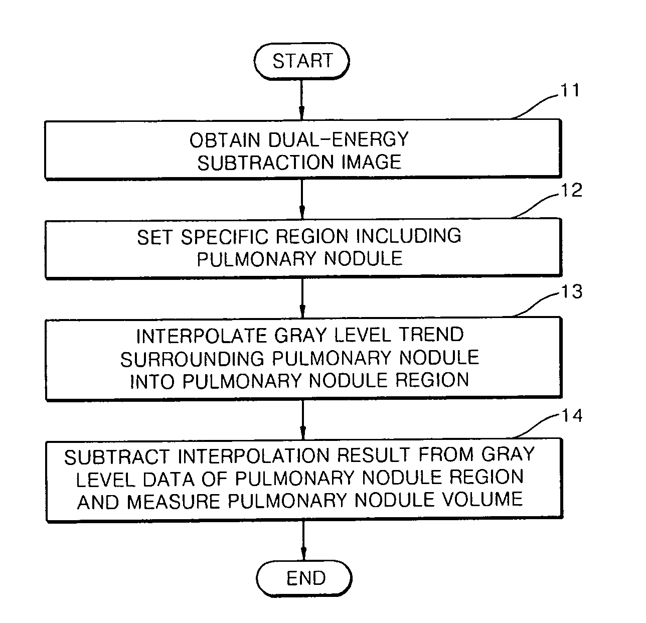
[0037] Hereinafter, the present invention will be described in detail by explaining embodiments of the invention with reference to the attached drawings. FIG. 1 is a flowchart illustrating a method of measuring a pulmonary nodule volume using a dual-energy subtraction image according to an exemplary embodiment of the present invention.

[0038] Referring to FIG. 1, according to the current embodiment of the present invention, a dual-energy subtraction image of a chest is obtained at first (process 11).

[0039] The dual-energy subtraction image can be obtained using a known dual-energy subtraction imaging device. The dual-energy subtraction imaging device can provide an approximately constant subtraction image with respect to the same part of a body, because the x-ray radiation conditions are stable. In general, the dual-energy subtraction imaging device sequentially irradiates two different x-rays of high and low energy respectively to a part of a body, obtains the high and low energy ...


PUM


Abstract

A method and a device for measuring a pulmonary nodule volume using a dual-energy subtraction image are provided. The method includes: (a) obtaining a dual-energy subtraction image of a chest; (b) setting a specific region including a pulmonary nodule region in the obtained dual-energy subtraction image; (c) interpolating a gray level trend of a region surrounding the pulmonary nodule region into the pulmonary nodule region using a fitting function; and (d) measuring the pulmonary nodule volume by subtracting the interpolation result from the gray level data of the pulmonary nodule region. Accordingly, when it is desired to measure the pulmonary nodule volume using the dual-energy subtraction image, information on the thickness of the pulmonary nodule is obtained from the characteristic of the dual-energy subtraction image, and the pulmonary nodule volume is accurately measured by correcting errors at the border of the pulmonary nodule region using the fitting function.

Description

CROSS-REFERENCE TO RELATED PATENT APPLICATION [0001] This application claims the benefit of Korean Patent Application No. 10-2005-0120026, filed on Dec. 8, 2005, and 10-2006-0042013, filed on May 10, 2006, in the Korean Intellectual Property Office, the disclosure of which is incorporated herein in its entirety by reference. BACKGROUND OF THE INVENTION [0002] 1. Field of the Invention [0003] The present invention relates to a method and a device for measuring pulmonary nodule volume using a dual-energy subtraction image. [0004] 2. Description of the Related Art [0005] Lung cancer is the most common cancer and the leading cause of cancer deaths around the world. More than one million three hundred thousand people die of lung cancer around the world every year. In South Korea, more than twelve thousand people have died of lung cancer in 2002, and the death rate from lung cancer has been reported to be the highest of all. A pulmonary nodule is commonly observed on a chest radiograph an...

Claims


Application Information

Patent Timeline
IPC IPC(8): G06K9/00
CPCG06K9/00G06K2209/053G06V2201/032G06T7/0014
Inventor LEE, SOOYEULLEE, JEONG WONKIM, SEUNGHWAN
Owner ELECTRONICS & TELECOMM RES INST
Features
  • R&D
  • Intellectual Property
  • Life Sciences
  • Materials
  • Tech Scout
  • Unparalleled Data Quality
  • Higher Quality Content
  • 60% Fewer Hallucinations
Learn More