

Implantable medical device coated with a bioactive agent
a bioactive agent and implantable technology, applied in the field of human and veterinary medical devices, can solve the problems of thrombosis and/or inflammatory response, particular problems in the delivery of water-soluble drugs, such as anti-sense drugs, and achieve the effect of inhibiting cellular proliferation and/or restenosis
- Summary
- Abstract
- Description
- Claims
- Application Information
AI Technical Summary
Benefits of technology
Problems solved by technology
Method used
Image

Examples
example 1
f a Water-Soluble Drug from a Stent Having a PLA and a PACA Overcoat
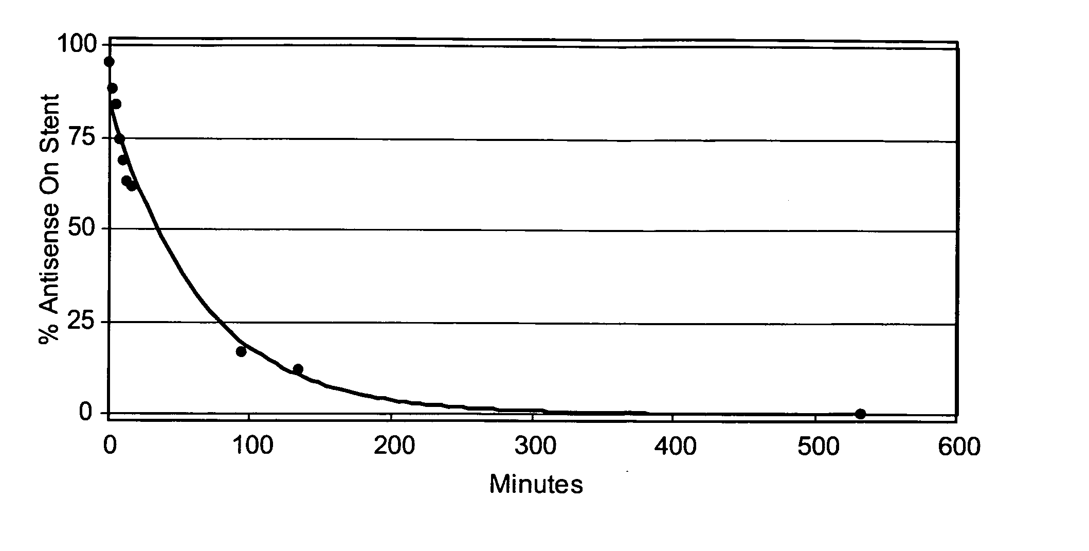
[0144] Three Zilver stents were coated with antisense drug and then with PLA using the protocol described in Example 2. Table 2 shows the amounts of antisense drug and PLA coated onto each stent. N-butyl cyanoacrylate monomer (NBCA) containing 10% Tri Butyl Citrate (plasticizer) and trace amounts of Butyl Hydroxyanisole and Sulphur Dioxide (stabilizers), (Glustitch, Inc., Washington 98281) was then applied to each stent using the method of Example 1. The coating solution contained 2.5 mg / ml NBCA in acetonitrile. The coating times were 40, 60 and 120 seconds for stents 1, 2 and 3 respectively. Table 2 shows the amount of NBCA coated onto the stents.
[0145] After coating, the elution time of the antisense drug was determined using the UV analysis method of Example 1. Table 2 shows the time taken for 90 percent of the coated antisense drug to elute from each stent.
TABLE 2Antisensen-butyl90% elutionStentDrug (μg)PLA (...
example 2
sing an Ultrasonic Coating Method
[0146] A mixture containing 4 mg / ml Resten-NG™ antisense drug and 8 mg / ml NBCA was prepared on 50:50 MeoH:CHCl3. The solution was loaded into a syringe mounted onto a syringe pump and connected to a tube that carried the solution to the ultrasonic nozzle (Sonotek, Milford, Conn. 06460). Air was purged from the solution line and the spray nozzle primed with the solution. The coating chamber was purged with nitrogen to attain an oxygen concentration of between 500 and 1500 ppm.
[0147] One end of Zilver stent was positioned on a mandrel. The nozzle was positioned about 100 mm from the stent and the stent was coated with about 419 μg of the mixture, while passing the nozzle over six loops of the stent surface. A power setting of 1.0 watts was used. The weight of mixture coated onto the stent was determined by weighing the stent before and after the coating procedure.
[0148] The stent was then over coated with a 4 mg / ml solution of PLA in dichloromethane ...
example 3
ith Two Barrier Layers
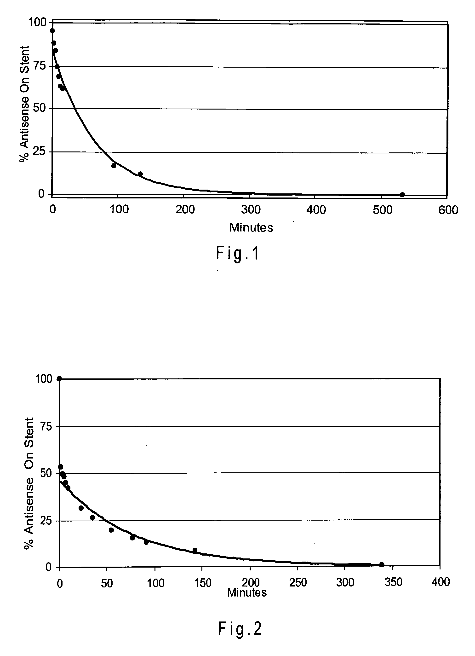
[0150] A Zilver stent was coated with about 320 μg of a mixture containing 4 mg / ml Resten-NG antisense drug and 5 mg / ml NBCA using the ultrasonic deposition method described in Example 2. A PLA overcoat (99 μg) was the applied, again using the ultrasonic method. Finally, 220 μg of NBCA was applied using the ultrasonic method.
[0151] The elution time of the antisense drug was determined using the UV analysis method of Example 1. FIG. 2 shows the elution of the antisense agent from the stent.
PUM
| Property | Measurement | Unit |
|---|---|---|
| Solubility (mass) | aaaaa | aaaaa |
| Solubility (mass) | aaaaa | aaaaa |
| Mass | aaaaa | aaaaa |
Abstract
Description
Claims
Application Information