Compact and durable encasement for a digital radiography detector

a detector and compact technology, applied in the field of medical imaging systems, can solve the problems of large, heavy, and difficult to obtain diagnostic images, and the detectors used in projection digital radiography are relatively large, and require substantial capital investmen

Active Publication Date: 2007-11-29
CARESTREAM HEALTH INC
View PDF14 Cites 134 Cited by
  • Summary
  • Abstract
  • Description
  • Claims
  • Application Information

AI Technical Summary

Problems solved by technology

The delay in obtaining a diagnostic image, the use of a chemical or thermal processor, and the difficulty in providing the radiographic film outside of the immediate medical facility, has resulted in the development of digital radiographic imaging systems.
The CR systems still result in a delay in obtaining a diagnostic image due to the necessity of scanning an exposed CR plate.
The solid-state, ionizing radiation based image detectors used in projection digital radiography today are relatively large, heavy, and expensive.
Additionally, a complete DR systems using thi

Method used


Image

  • Compact and durable encasement for a digital radiography detector
  • Compact and durable encasement for a digital radiography detector
  • Compact and durable encasement for a digital radiography detector

Examples


Embodiment Construction

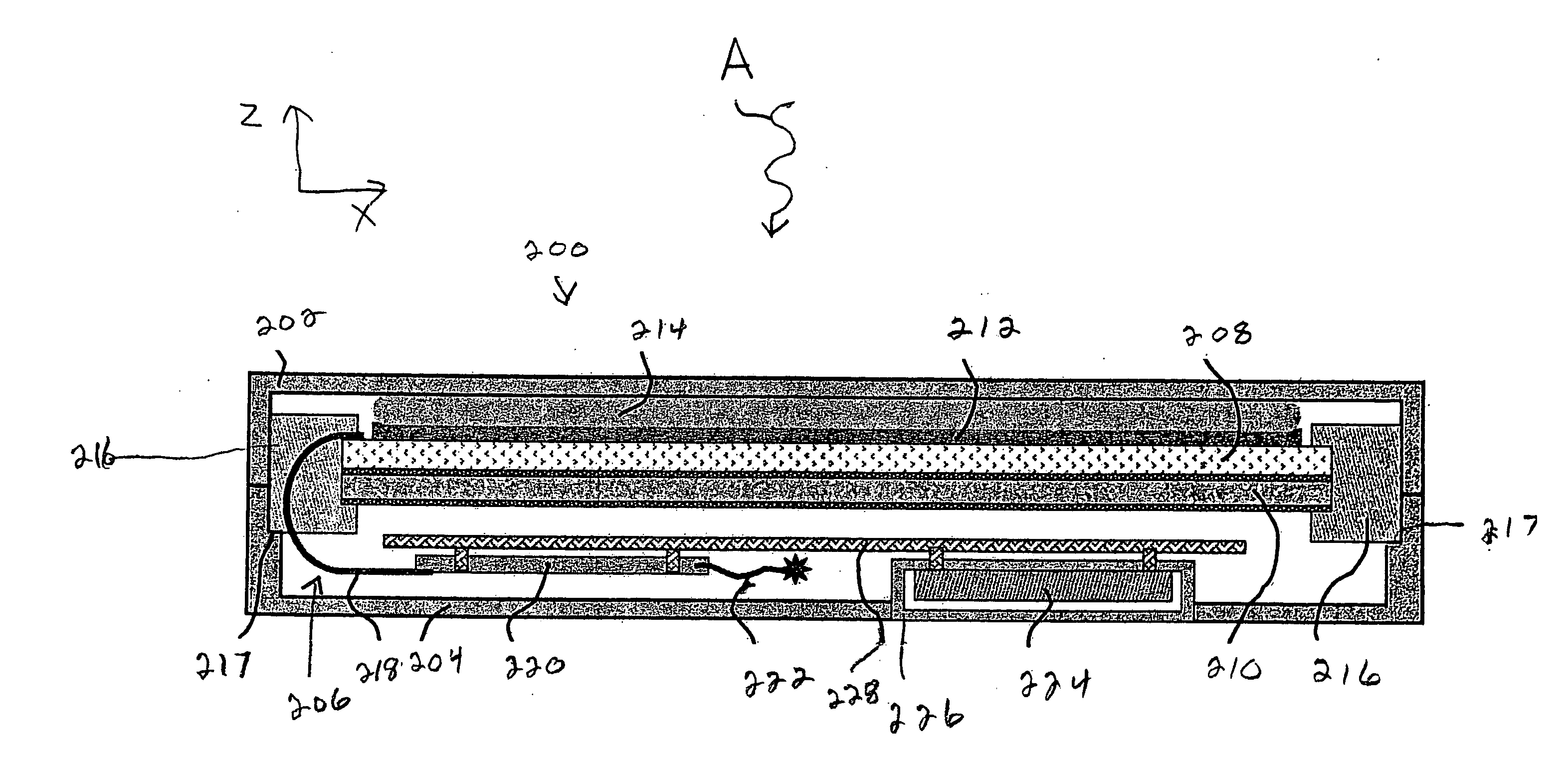
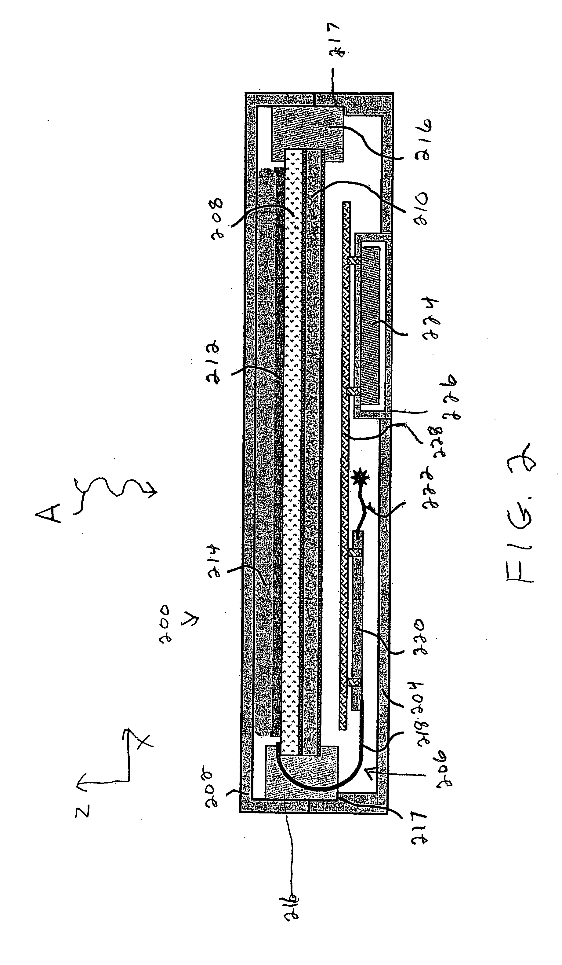
[0043]The following is a detailed description of the preferred embodiments of the invention, reference being made to the drawings in which the same reference numerals identify the same elements of structure in each of the several figures.

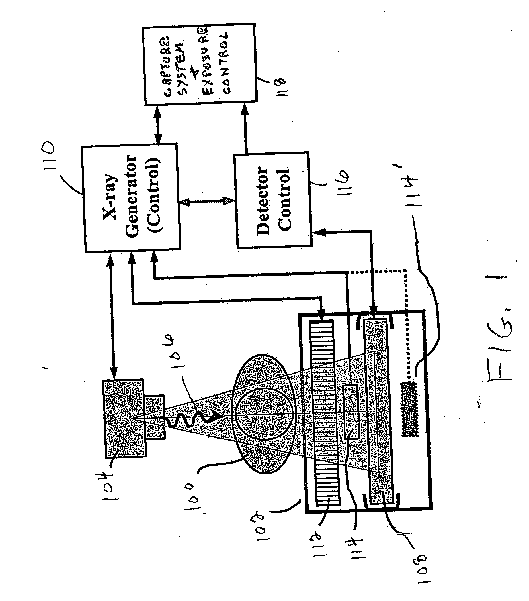
[0044]Referring now to FIG. 1, there is shown diagrammatically typical projection x-ray equipment used in an x-ray examination room. As shown, a patient 100 is positioned on a support 102. An x-ray source 104 projects x-rays 106 through a body part of patient 100 to form a radiographic image of the body part which is detected by a digital detector housed in radiography cassette 108 mounted in support 102. X-ray source 104 is activated and controlled by x-ray generator and control 110. Support (Bucky) 102 can also house an antiscatter grid 112, an auto exposure control sensor 114, 114′ (located above the radiography cassette for general radiography and below the radiography cassette for mammography). Detector control 116 is linked to the digital dete...


PUM


Abstract

A digital radiography detector includes a housing having first and second spaced planar members and four side walls defining a cavity. A radiographic image detector assembly is mounted within the cavity for converting a radiographic image to an electronic radiographic image. The detector assembly includes a detector array mounted on a stiffener. A shock absorbing elastomer assembly is located within the cavity for absorbing shock to the detector array/stiffener in directions perpendicular to and parallel to the detector array/stiffener.

Description

FIELD OF THE INVENTION[0001]This invention relates in general to medical imaging systems which use digital radiography detectors, and more particularly to a compact and durable encasement or housing for a digital radiography detector.BACKGROUND OF THE INVENTION[0002]Traditional film-screen radiography has been used as a medical imaging diagnostic system for many years. X-rays are projected through a patient's body part to form a latent radiographic image on film contained in a cassette. The film is then be chemically or thermally processed to produce a visual radiographic image which can be used by a health care professional for diagnostic purposes. The delay in obtaining a diagnostic image, the use of a chemical or thermal processor, and the difficulty in providing the radiographic film outside of the immediate medical facility, has resulted in the development of digital radiographic imaging systems. Computed radiography (CR) digital systems have been developed in recent years that...

Claims


Application Information

Patent Timeline
IPC IPC(8): G01T1/20
CPCG01T1/20
Inventor JADRICH, BRADLEY S.PETER, TIMOTHY A.DHURJATY, SREERAMHAWVER, JEFFERY R.BEDZYK, MARK D.
Owner CARESTREAM HEALTH INC
Why Eureka
Try Eureka