Integrated handle assembly for anchor delivery system

a handle and anchor technology, applied in the field of medical devices and methods, can solve the problems of reducing the quality of life of patients, laborious surgical dissection, and reducing the volume of prostate glands, so as to facilitate the testing of the effectiveness of positioning of an anchor assembly, facilitate healing, and minimize infection risk

Active Publication Date: 2007-11-29
TELEFLEX LIFE SCI LTD
View PDF99 Cites 80 Cited by
  • Summary
  • Abstract
  • Description
  • Claims
  • Application Information

AI Technical Summary

Benefits of technology

[0040] The present invention also contemplates a reversible procedure as well as an anchor assembly with sufficient visibility when viewed ultrasonically. In one aspect, the implant procedure is reversible by severing a connector of an anchor assembly and removing an anchor of the anchor assembly such as by so removing a proximally placed anchor previously implanted in an urethra. Moreover, the anchor assemblies can be formed of structures facilitating ultrasound viewing.
[0042] In another aspect, structure of the anchor assembly is designed to invaginate within or complement tissue anatomy to thereby facilitate healing and minimize infection risk. Moreover, the anchor delivery device includes structure to form desired angles between an extended position of the needle assembly relative to the device. Additionally, it is contemplated that a distal end portion of the anchor delivery device be configured to facilitate the testing of the effectiveness of positioning of an anchor assembly. In this regard, the distal end portion is configured in a manner to mimic the effect a second anchor member will have prior to its implantation.

Problems solved by technology

In many of these procedures, surgical incisions are made in the body and laborious surgical dissection is performed to access and expose the affected tissues or anatomical structures.
This combination of increased urethral resistance to urine flow and stiffness and hypertrophy of urinary bladder walls leads to a variety of lower urinary tract symptoms (LUTS) that may severely reduce the patient's quality of life.
Although BPH is rarely life threatening, it can lead to numerous clinical conditions including urinary retention, renal insufficiency, recurrent urinary tract infection, incontinence, hematuria, and bladder stones.
Thus 5-alpha-reductase inhibitors cause regression of epithelial cells in the prostate gland and hence reduce the volume of the prostate gland which in turn reduces the severity of the symptoms.
This generates heat which in turn destroys the prostate tissue.
This creates coagulation defects in the lateral lobes.
This heats the middle lobe and causes laser induced necrosis of the tissue around the distal tip.
This causes tissue necrosis in several regions of the prostate gland which in turn causes the prostate gland to shrink.
Although existing treatments provide some relief to the patient from symptoms of BPH, they have disadvantages.
Surgical treatments of BPH carry a risk of complications including erectile dysfunction; retrograde ejaculation; urinary incontinence; complications related to anesthesia; damage to the penis or urethra, need for a repeat surgery etc.
Even TURP, which is the gold standard in treatment of BPH, carries a high risk of complications.
In fact, symptoms are typically worse for these therapies that heat or cook tissue, because of the swelling and necrosis that occurs in the initial weeks following the procedures.
Prostatic stents often offer more immediate relief from obstruction but are now rarely used because of high adverse effect rates.
Overgrowth of tissue through the stent and complex stent geometries have made their removal quite difficult and invasive.
Thus the most effective current methods of treating BPH carry a high risk of adverse effects.
These methods and devices either require general or spinal anesthesia or have potential adverse effects that dictate that the procedures be performed in a surgical operating room, followed by a hospital stay for the patient.
While several of these procedures can be conducted with local analgesia in an office setting, the patient does not experience immediate relief and in fact often experiences worse symptoms for weeks after the procedure until the body begins to heal.
While drug therapies are easy to administer, the results are suboptimal, take significant time to take effect, and often entail undesired side effects.
The severity of UI varies and, in severe cases, the disorder can be totally debilitating, keeping the patient largely homebound.
The surgeries used to treat UI are generally associated with significant discomfort as the incisions heal and may require a Foley or supra-pubic urinary catheter to remain in place for at least several days following the surgery.

Method used


Image

  • Integrated handle assembly for anchor delivery system
  • Integrated handle assembly for anchor delivery system
  • Integrated handle assembly for anchor delivery system

Examples


Embodiment Construction

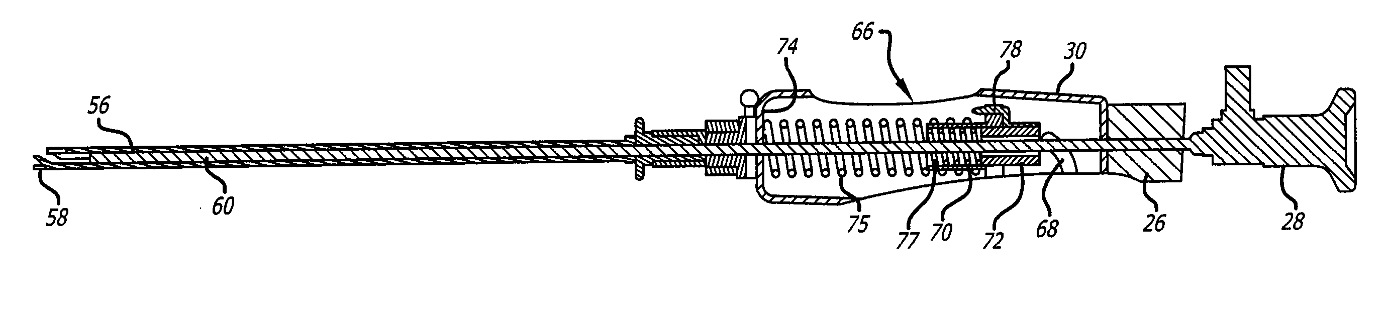
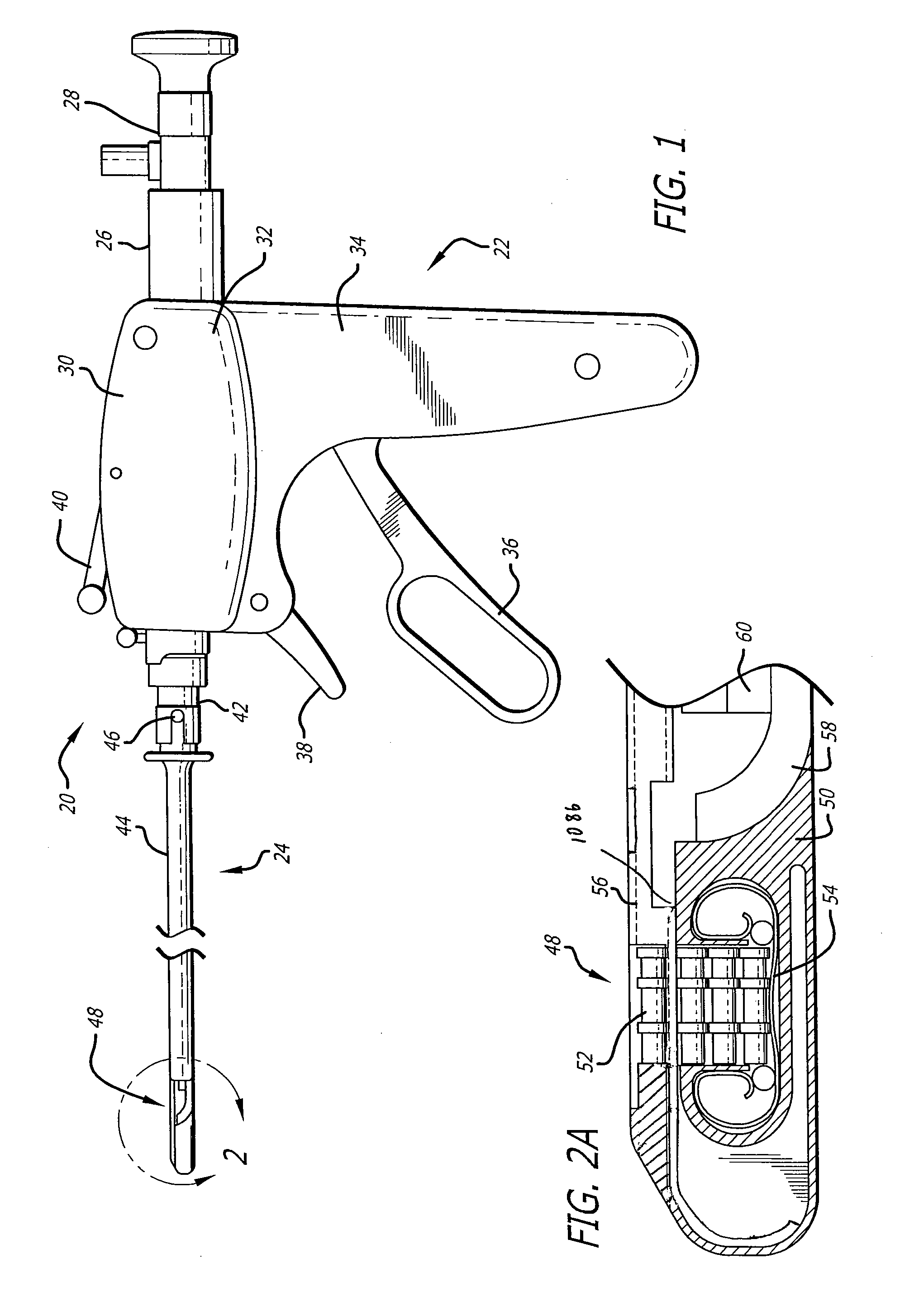
[0175] Turning now to the figures, which are provided by way of example and not limitation, the present invention is embodied in a device configured to deliver anchor assemblies within a patient's body. As stated, the present invention can be employed for various medical purposes including but not limited to retracting, lifting, compressing, supporting or repositioning tissues, organs, anatomical structures, grafts or other material found within a patient's body. Such tissue manipulation is intended to facilitate the treatment of diseases or disorders. Moreover, the disclosed invention has applications in cosmetic or reconstruction purposes or in areas relating the development or research of medical treatments.

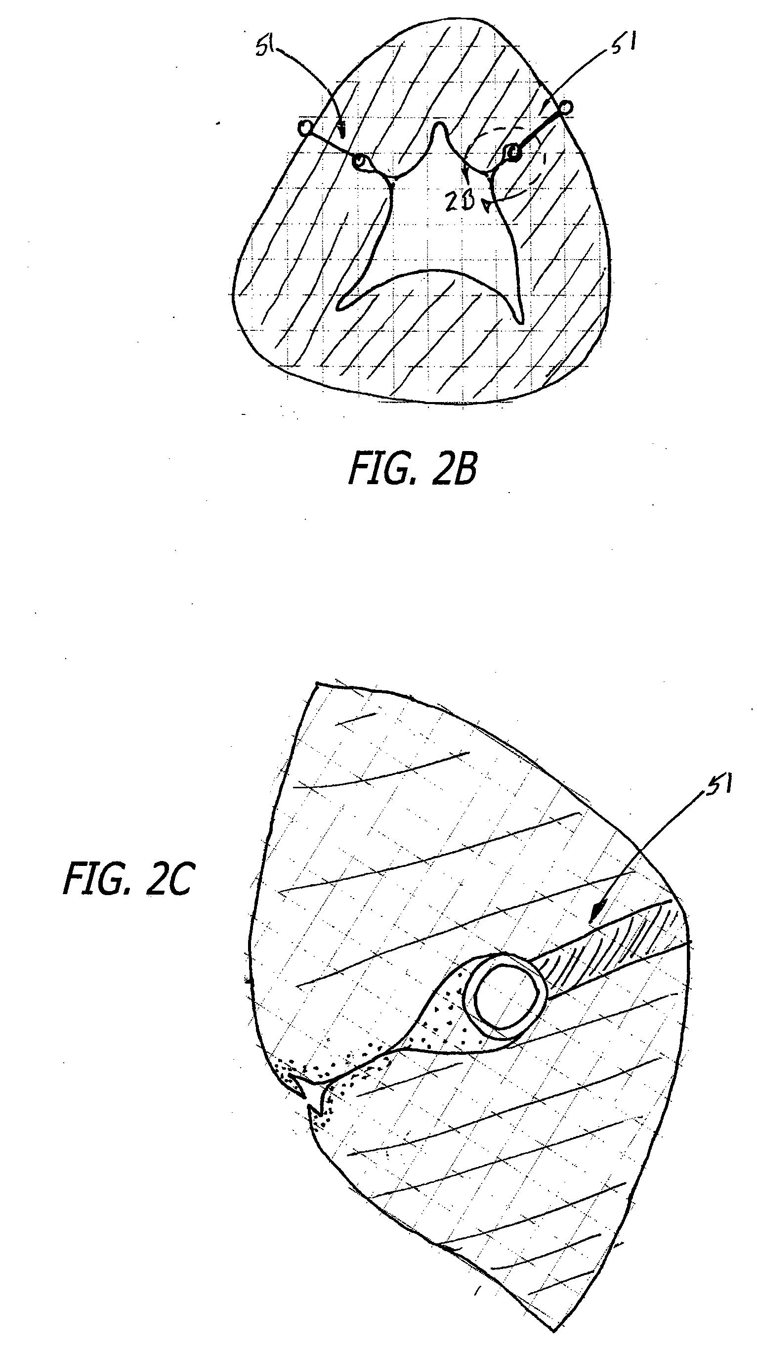
[0176] In one particular aspect, the anchor assembly of the present invention is contemplated to be formed of a structure which is visible by ultrasound. Accordingly, the anchor assembly can be viewed during ultrasonic body scans such as during normal trans-rectal ultrasound ...


PUM


Abstract

Intergrated systems and associated method for manipulating tissues and anatomical or other structures in medical application for the purpose of treating diseases or disorders or other purposes. In one aspect, the system includes a delivery device configured to deploy and implant anchor device for such purposes.

Description

CROSS-REFERENCES TO RELATED APPLICATIONS [0001] This application is a continuation-in-part of copending U.S. patent application Ser. No. 11 / 492,690, entitled Apparatus and Method for Manipulating or Retracting Tissue and Anatomical Structure, filed on Jul. 24, 2006, which is a continuation-in-part of copending U.S. patent application Ser. No. 11 / 318,246, entitled Devices, Systems and Methods for Retracting, Lifting, Compressing, Supporting or Repositioning Tissues or Anatomical Structures, filed on Dec. 22, 2005, which is a continuation-in-part of copending U.S. patent application Ser. No. 11 / 134,870 entitled Devices, Systems and Methods for Treating Benign Prostatic Hyperplasia and Other Conditions, filed on May 20, 2005, the entire disclosures of which are expressly incorporated herein by reference.FIELD OF THE INVENTION [0002] The present invention relates generally to medical devices and methods, and more particularly to integrated systems and associated methods for manipulating...

Claims


Application Information

Patent Timeline
Patent Type & Authority Applications(United States)
IPC IPC(8): A61B17/128
CPCA61B17/0218A61F2/0045A61B17/0469A61B17/0482A61B17/0487A61B17/0625A61B17/32A61B17/3468A61B17/3478A61B17/42A61B2017/00022A61B2017/00274A61B2017/0046A61B2017/00792A61B2017/00805A61B2017/0404A61B2017/0409A61B2017/0417A61B2017/0419A61B2017/045A61B2017/0454A61B2017/0464A61B2017/0488A61B2017/06052A61B2017/06176A61B2017/0688A61B2018/00547A61B17/0401A61B17/00234A61B17/0467A61B17/10A61B2017/00796A61F2/0022A61F2/0063
Inventor CATANESE, JOSEPH IIIMCLEAN, MATTLAMSON, THEODORE C.NAGPURKAR, AMIKMERRICK, DANIELMAKOWER, JOSHUAVIDAL, CLAUDEREDMOND, RUSSELL J.COLLINSON, MICHAELBARHAM, MITCHELL C.WEST, SCOTT
Owner TELEFLEX LIFE SCI LTD
Why Eureka
Try Eureka