[0008]The object of the invention is to improve a catheter of the type mentioned above such that it allows removal of tissue in a hollow organ in a protective manner and its function is able to be observed easily during use, and also that it makes it possible for a vessel blockage to be completely treated by a one-off introduction of the inventive catheter.
[0009]To resolve this problem, the invention makes provision, with a catheter of the type mentioned at the start, for a
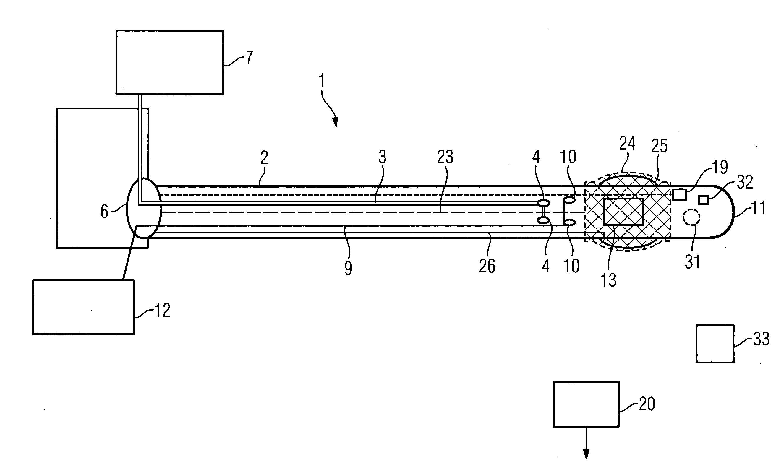
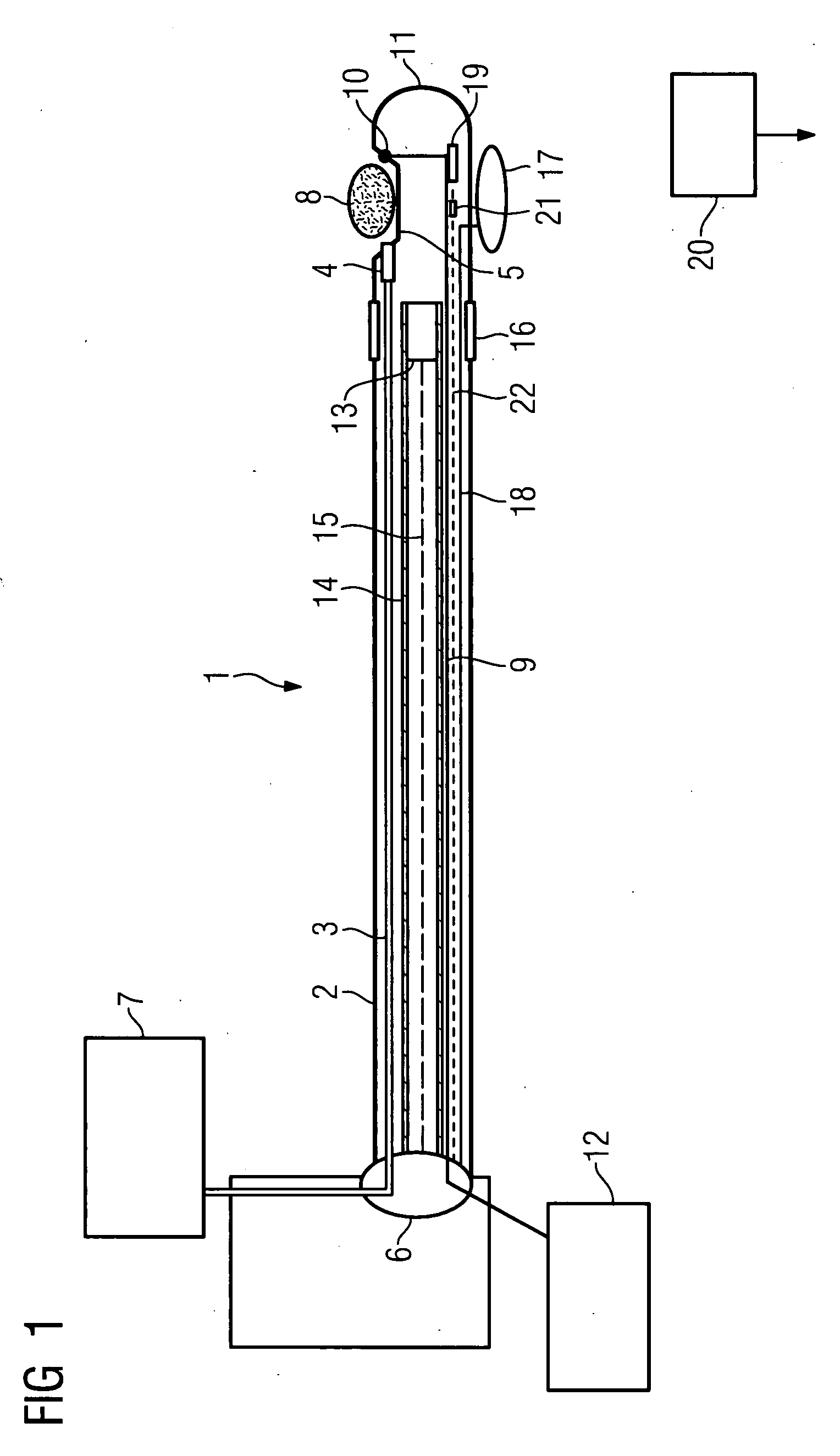
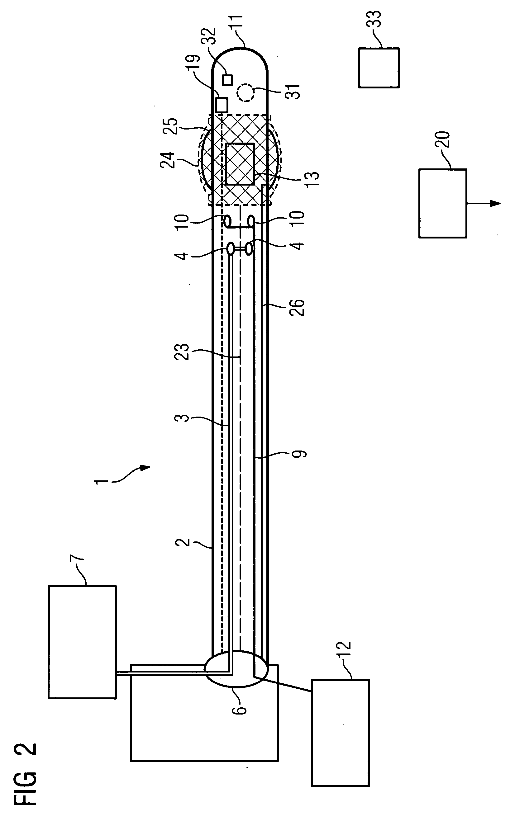
stent to be provided in the area of the catheter tip, which can be expanded to place it via a
balloon located underneath it which is able to be inflated via a further lumen. This metallic
stent, made of stainless steel for example, can also be covered by a medical or therapeutic layer respectively (
Drug Eluting
Stent). Alternatively it can consist of a bioresorbable material which dissolves within a predetermined time. The Integration of such a stents thus enables a vessel blockage to be completely treated by a single
insertion of the inventive catheter.
[0010]The outstanding feature of the inventive catheter is an imaging facility integrated into the tip, the recording area of which is directed towards the area of tissue treated with the emitted jet of fluid. This allows continuous observation both during the treatment and also beforehand and afterwards. The user also has the option on the one hand of using the integrated
image recording device to orient themselves in situ, after obtaining the local circumstances directly via the image recording device. He can continuously check the
removal procedure while undertaking it and can also immediately check the success of the treatment after it has been undertaken and make any necessary adjustments. An exact assignment of the
stenosis to the position of the treatment area of the catheter is thus a simple matter.
[0013]Expediently with this embodiment a further lumen for supplying a contrast fluid to a further outlet opening provided in the area of the catheter tip is provided, via which the contrast fluid is emitted into the imaging area. Such a
contrast medium, e.g.
sulfur hexafluoride, forms temporary gas bubbles in the bloodstream and changes the reflection properties of the
ultrasound signals, which leads to an improvement in
image quality. Optionally a further lumen can be provided for supplying an x-
ray contrast medium to enable an x-
ray check to be undertaken in parallel if required.
[0020]In a further development one guiding wire or a number of guiding wires to make possible an explicit bending of the catheter tip for an easier movement of the catheter tip through the vessel or such like can be provided, with the said guiding wires being routed to an external location and operated from there. In addition a definable tip bending in their direction can be achieved so that it is significantly easier to maneuver around any vessel bends etc.
[0023]There is also the option of manufacturing the catheter or the shell respectively etc. from materials which do not influence the image recording, i.e. of materials which for example shield against magnetic fields in the case of an IVMRI image recording device etc. The catheter can also be provided with a
coating which reduces the
frictional resistance during guidance. This
coating can be made of
silicon for example. Finally there is the option of decoupling the connections for the physiological sensors etc via a suitable
electrical isolation from any mains
voltage, in order not to endanger the patient. Optical decoupling is especially advantageous here. There is also the option of providing the catheter with a detection means, for example an RFID
transponder via which the catheter can be exactly defined and which corresponds with an appropriate control device etc. for presetting the entire
system in relation to the application of the catheter.

