Methods for displaying a location of a point of interest on a 3-d model of an anatomical region

a technology of anatomical region and location, applied in the field of methods for displaying the location of a point of interest on a 3d model of anatomical region, can solve the problem that the point of interest identified on a 2-d image cannot be automatically identified in a 3-d model of the anatomical region

Active Publication Date: 2008-06-26
APN HEALTH +1
View PDF10 Cites 37 Cited by
  • Summary
  • Abstract
  • Description
  • Claims
  • Application Information

AI Technical Summary

Problems solved by technology

However, a drawback associated with the fluoroscopy system and the CT system, is that a point of interest identified on a 2-D image cannot be automatically identified in a 3-D model of the anatomical region.

Method used


Image

  • Methods for displaying a location of a point of interest on a 3-d model of an anatomical region
  • Methods for displaying a location of a point of interest on a 3-d model of an anatomical region
  • Methods for displaying a location of a point of interest on a 3-d model of an anatomical region

Examples


Embodiment Construction

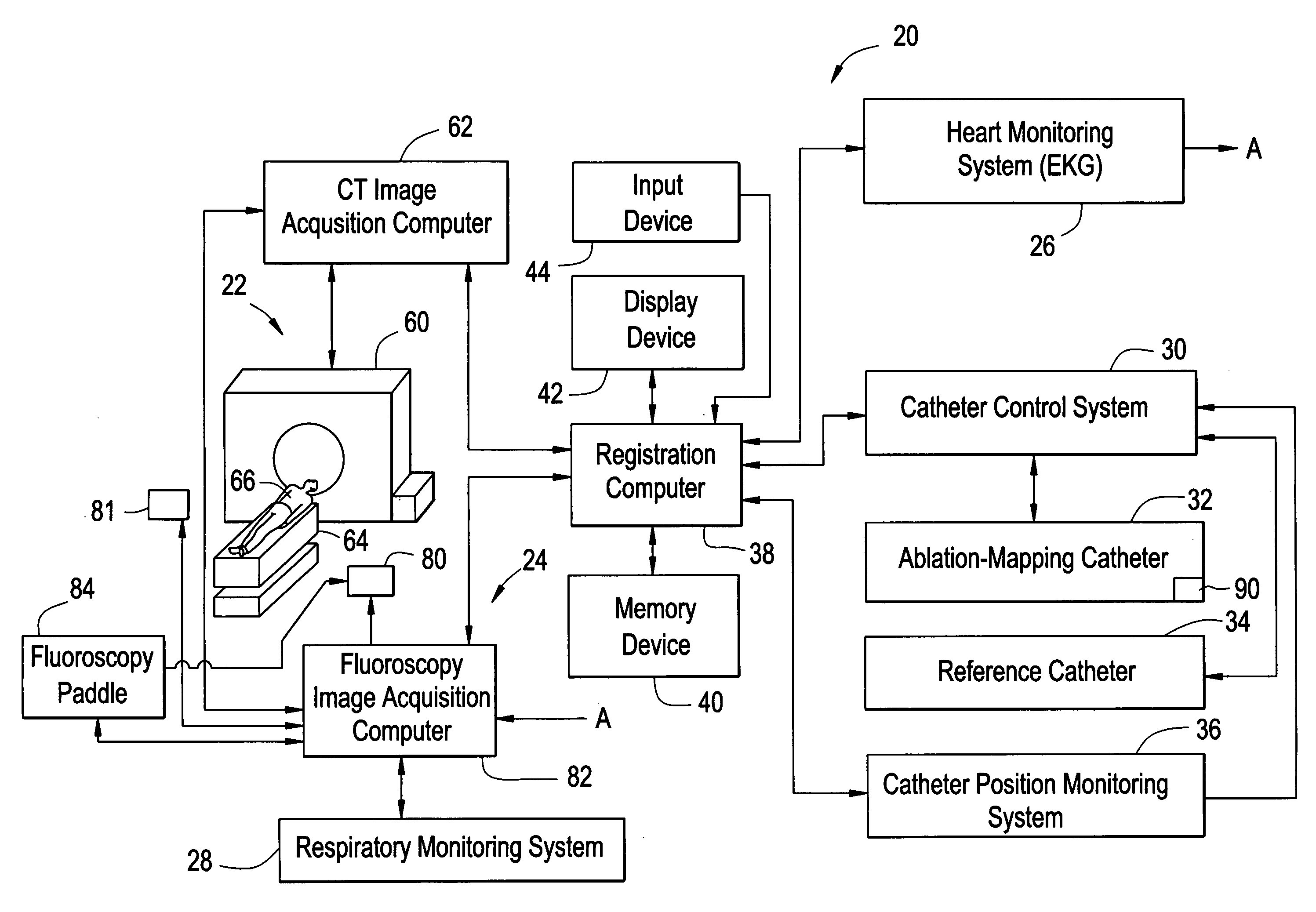
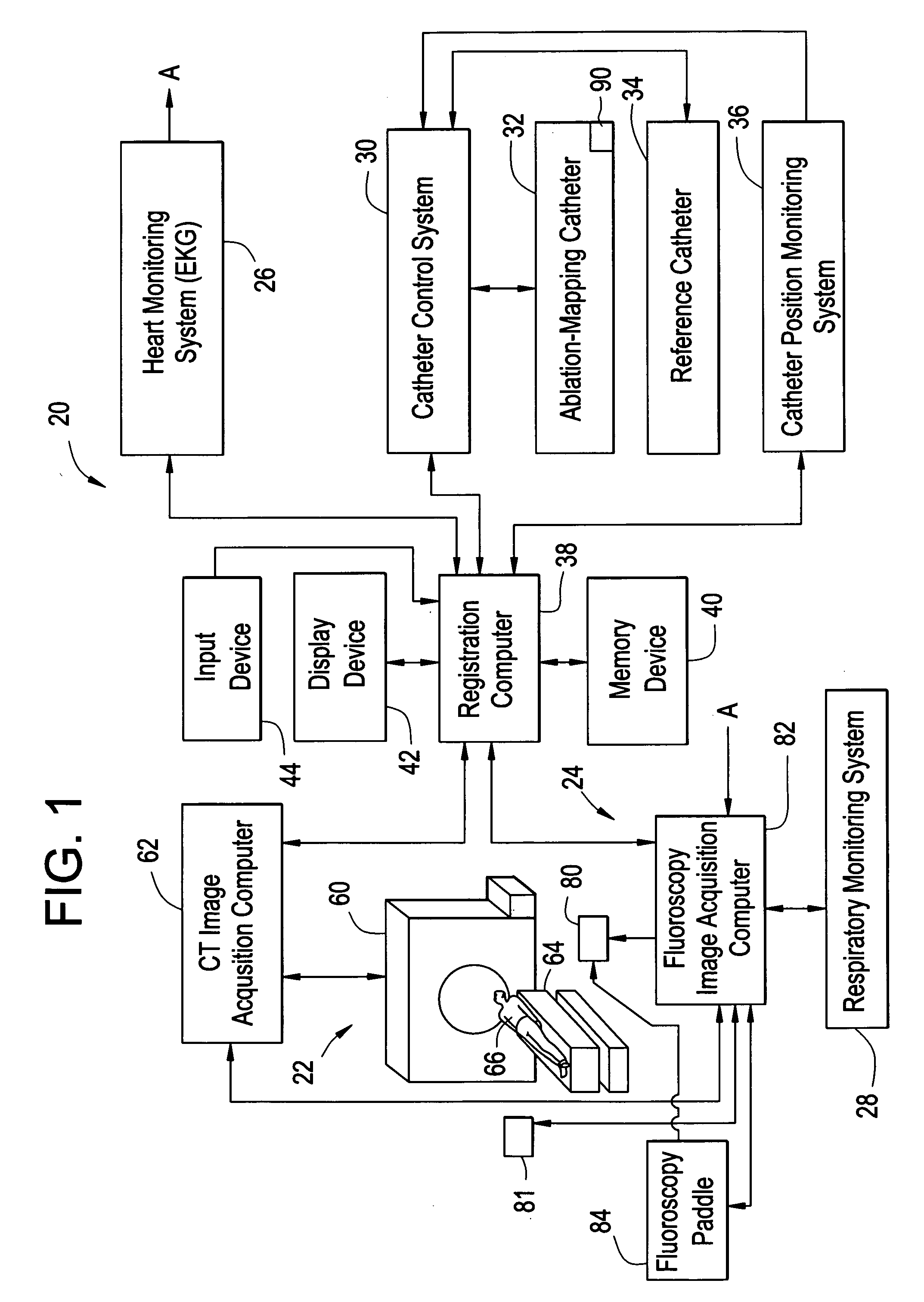
[0034]Referring to FIG. 1, a schematic of a system 20 for displaying a location of a point of interest on a 3-D model of an anatomical region and for generating a registered image in accordance with an exemplary embodiment is illustrated. The system 20 includes a computed tomography (CT) image acquisition system 22, a fluoroscopy image acquisition system 24, a heart monitoring system 26, a respiratory monitoring system 28, a catheter control system 30, and ablation-mapping catheter 32, a reference catheter 34, a catheter position monitoring system 36, a registration computer 38, a memory device 40, a display device 42, and an input device 44.

[0035]The CT image acquisition system 22 is provided to generate a 3-D model of the anatomical region of a person 66. The CT image acquisition system 22 includes a CT scanning device 60, a CT image acquisition computer 62, and a table 64. The CT scanning device 60 generates scanning data of the anatomical region of the person 66 who is disposed ...


PUM


Abstract

Methods for displaying a location of a point of interest on a 3-D model of an anatomical region of a person are provided. In one exemplary embodiment, the method includes generating first and second 2-D images of the anatomical region utilizing an image acquisition system when an X-ray source of the image acquisition system is disposed at first and second positions, respectively, in a 3-D coordinate system of the image acquisition system. The method further includes selecting first and second points in the first and second 2-D images, respectively. The method further includes utilizing a triangulation technique utilizing the first and second points to determine a point of interest in a 3-D coordinate system of the 3-D model of the anatomical region.

Description

CROSS REFERENCE TO RELATED APPLICATION[0001]The application claims the benefit of U.S. Provisional application, Ser. No. 60 / 855,115, filed Oct. 30, 2006, the contents of which are incorporated herein by reference thereto.BACKGROUND OF THE INVENTION[0002]A fluoroscopy system has been utilized to generate a 2-D image of an anatomical region. Further, a CT system has been utilized to generate a 3-D model an anatomical region. However, a drawback associated with the fluoroscopy system and the CT system, is that a point of interest identified on a 2-D image cannot be automatically identified in a 3-D model of the anatomical region.[0003]The inventors herein have recognized a need for a method for displaying a location of a point of interest on a 3-D model of an anatomical region, utilizing at least one point on at least one 2-D image of the anatomical region.BRIEF DESCRIPTION OF THE INVENTION[0004]A method for displaying a location of a point of interest on a 3-D model of an anatomical r...

Claims


Application Information

Patent Timeline
Patent Type & Authority Applications(United States)
IPC IPC(8): A61B6/00
CPCA61B6/032A61B6/541A61B6/5235A61B6/487A61B6/503Y10S128/922
Inventor VAILLANT, REGISSOUBELET, ELISABETHSRA, JASBIR SINGH
Owner APN HEALTH
Why Eureka
Try Eureka