

Methods and Apparatus For Performing Enhanced Ultrasound Diagnostic Breast Imaging
a breast imaging and ultrasound technology, applied in the field of medical diagnostic imaging systems, can solve the problems of cumbersome use of conformal gel pads or water bags, inability to image breast volume, and inability to achieve the full volume of breast ultrasound,
- Summary
- Abstract
- Description
- Claims
- Application Information
AI Technical Summary
Benefits of technology
Problems solved by technology
Method used
Image


Examples
Embodiment Construction
[0015]In the figures, like reference numerals refer to like elements. In addition, it is to be noted that the figures are not drawn to scale.
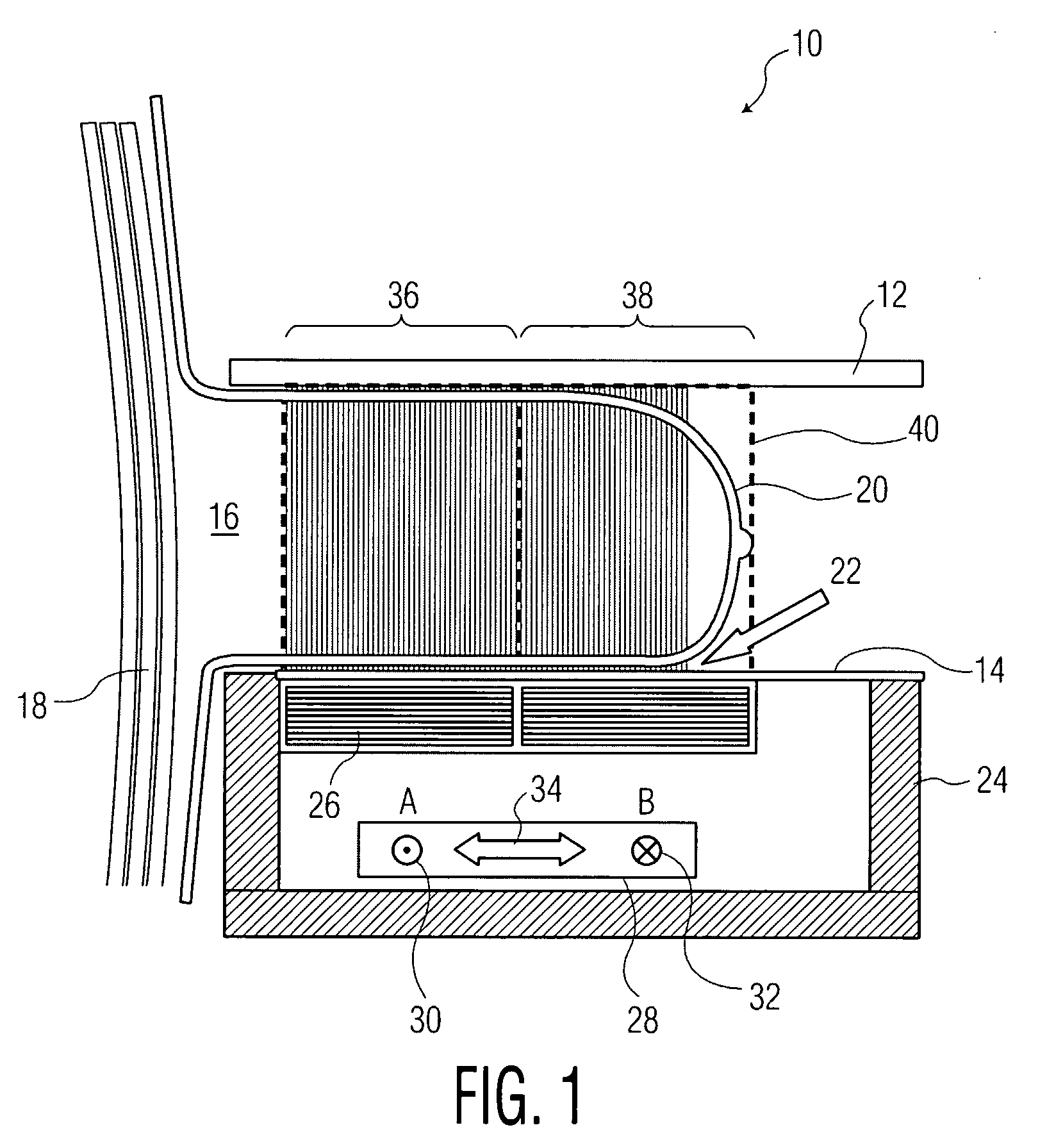
[0016]FIG. 1 is a cross-sectional view of a portion of an ultrasound diagnostic breast imaging system that uses a two pass linear scan for obtaining a conventional, non-steered rectangular volumetric image. That is, the embodiment of FIG. 1 uses non-steered rectangular images to acquire 3D volumes. As a result, the two-pass (A and B) 3D scan with conventional, non-steered rectangular image frames fails to image the curved area near the nipple and the tissue adjacent to the chest wall, as discussed further below.
[0017]As shown in FIG. 1, the ultrasound diagnostic breast imaging system 10 includes a first compression plate 12 and a second compression plate 14. The first and second plates are configured for receiving a breast 16 and further adapted for compressing the breast between the first and second plates. The breast 16 extends from a chest w...
PUM
 Abstract
Description
Claims
Application Information