Biopsy device with multiple cutters

Inactive Publication Date: 2008-12-04
BOSTON SCI SCIMED INC
View PDF19 Cites 29 Cited by
  • Summary
  • Abstract
  • Description
  • Claims
  • Application Information

AI Technical Summary

Benefits of technology

[0007]In one embodiment, each cutter has a semi-circular cross-section. In an optional embodiment, the tissue sampling device comprises a firing mechanism (e.g., a pneumatic or spring-loaded mechanism) configured to rapidly advance each cutter towards the distal end of the needle and across the respective aperture to sever the prolapsed tissue. In another optional

Problems solved by technology

In addition to increased patient discomfort, the reinsertion of

Method used


Image

  • Biopsy device with multiple cutters
  • Biopsy device with multiple cutters
  • Biopsy device with multiple cutters

Examples


Embodiment Construction

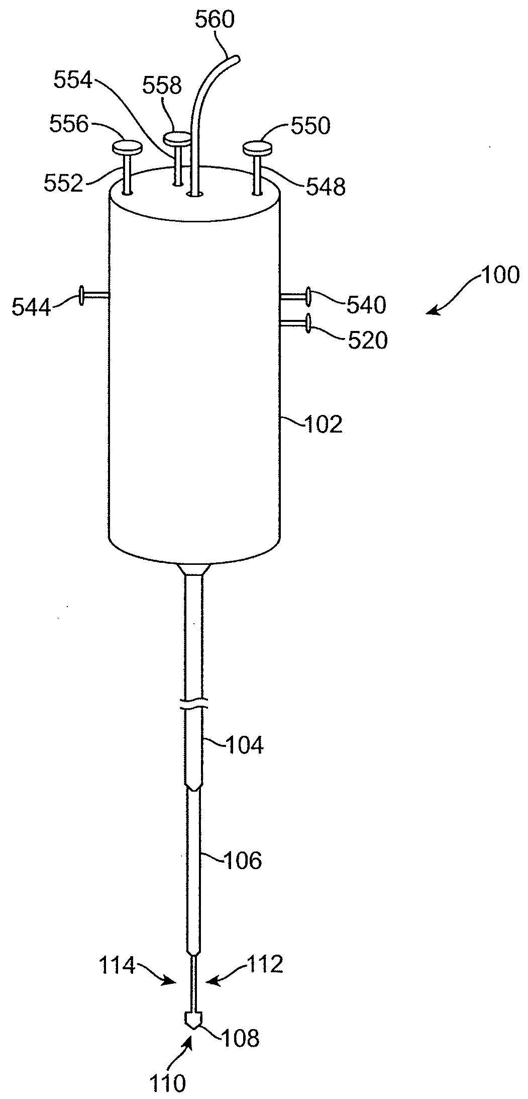
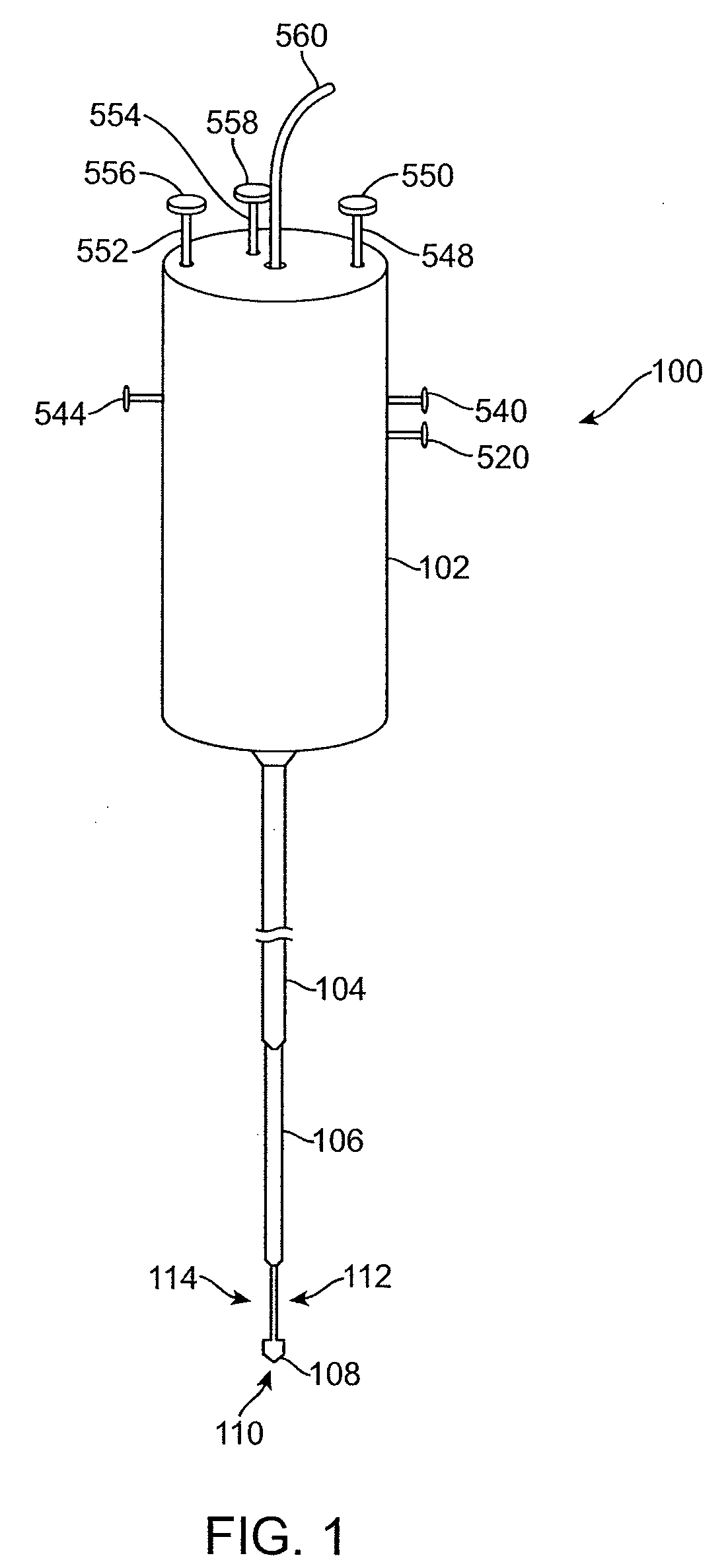
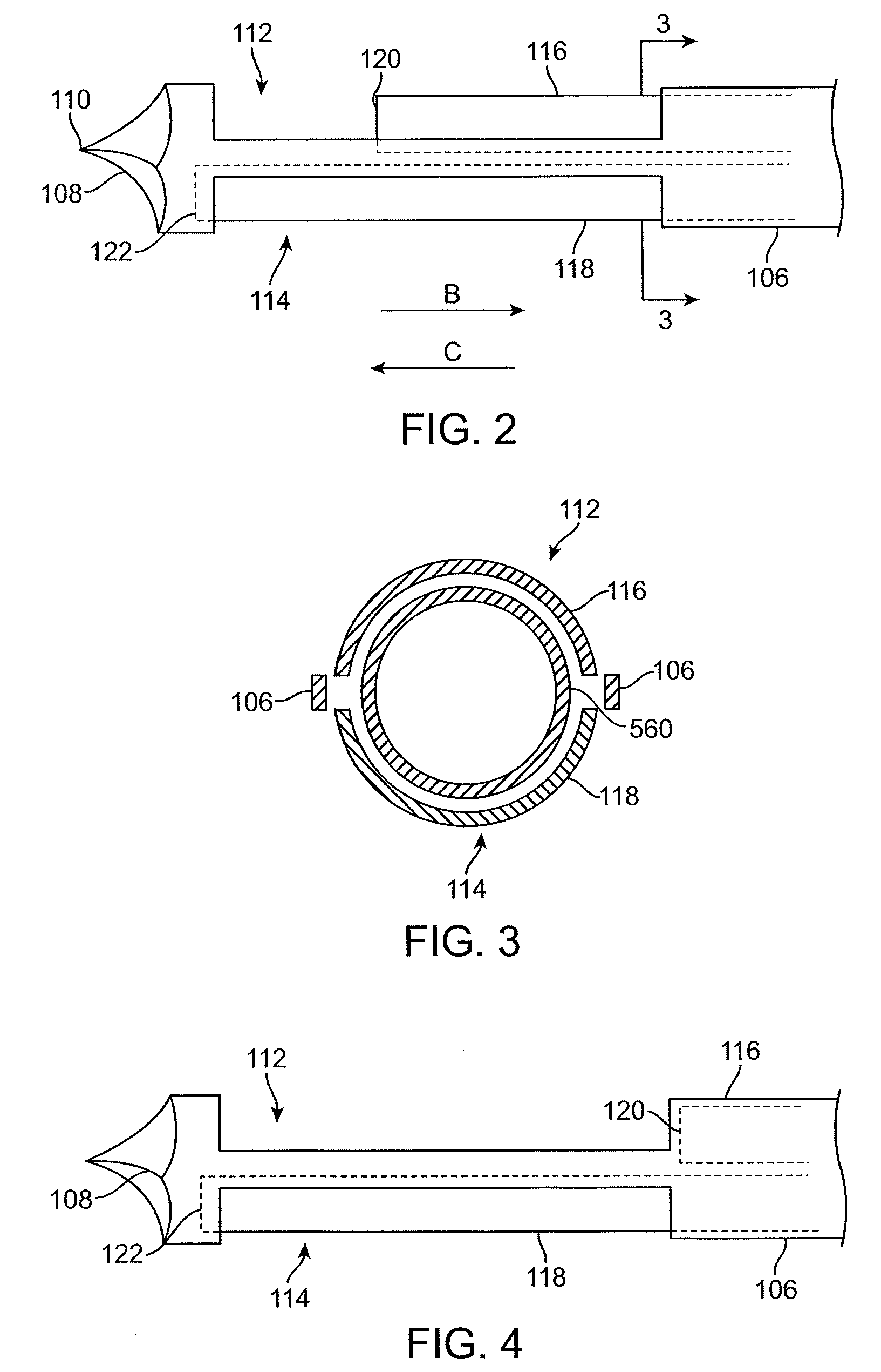
[0025]In one embodiment, a biopsy device having a needle is provided. The needle disclosed herein comprises at least two apertures, each of which interacts with a cutter, and each cutter operates independently. By using the needle disclosed herein, a surgeon or other medical professional can obtain two or more biopsy specimen without the need to reinsert the needle. The number of device manipulation, and therefore, the time needed to complete the procedure, is thus cut down significantly. For purposes of illustration, the use of the embodiments of the needle disclosed herein is described with examples of various biopsy devices. However, those of ordinary skill in the art recognize that the embodiments of the needle disclosed herein can be used with any of the known biopsy devices, for example, those disclosed in U.S. Pat. Nos. 5,989,196, 5,368,045, 5,573,008, 5,823,971, 6,165,136, 6,273,861, and 7,001,341, each of which is incorporated by reference herein in its entirety, including ...


PUM


Abstract

A tissue sampling device comprises an elongated tube having a distal end with at least two apertures. The device further comprises at least two cutters disposed within the elongated tube, wherein each cutter is configured to be independently displaced longitudinally within the elongated tube and across a respective one of the apertures to sever tissue prolapsed within the respective aperture. A method comprises inserting the device in tissue. The method further comprises receiving a first prolapsed portion of the tissue into the first aperture, longitudinally displacing the first cutter across the first aperture to sever the first prolapsed tissue portion within the first aperture, receiving a second prolapsed portion of the tissue into the second aperture, and longitudinally displacing the second cutter independently from the first cutter across the second aperture to sever the second prolapsed tissue portion within the second aperture.

Description

RELATED APPLICATION DATA[0001]The present application claims the benefit under 35 U.S.C. § 119 to U.S. Provisional Patent Application Ser. No. 60 / 940,636, filed May 29, 2007. The foregoing application is incorporated by reference into the present application in its entirety for all purposes.FIELD OF THE INVENTION[0002]The present invention is in the field of devices to obtain biopsy samples.BACKGROUND[0003]The diagnosis and treatment of patients with cancerous tumors, premalignant conditions, and other disorders has long been an area of intense interest in the medical community. Non-invasive methods for examining tissue and, more particularly, breast tissue include palpation, X-ray imaging, MRI imaging, CT imaging, and ultrasound imaging. When a physician suspects that tissue may contain cancerous cells, a biopsy may be done using either an open procedure or in a percutaneous procedure. In an open procedure, a scalpel is used by the surgeon to create an incision to provide direct vi...

Claims


Application Information

Patent Timeline
IPC IPC(8): A61B10/02
CPCA61B10/0275A61B10/0283A61B2010/0208A61B2010/0225A61B2017/00539A61B2017/00544
Inventor MCINTYRE, JON T.
Owner BOSTON SCI SCIMED INC
Why Eureka
Try Eureka