Method and a device for simplifying a diagnostic assessment of a mechanically ventilated patient

a technology for mechanical ventilation and diagnostic assessment, which is applied in the field of methods and devices for simplifying the diagnostic assessment of mechanical ventilation patients, can solve the problems that mechanical ventilation may damage the lungs of patients, and studies have yet to show whether the representation remains intuitively perceptible and usable, so as to simplify the diagnostic assessment and simplify the parameter assessmen

Inactive Publication Date: 2009-01-22
HAMILTON MEDICAL AG
View PDF9 Cites 115 Cited by
  • Summary
  • Abstract
  • Description
  • Claims
  • Application Information

AI Technical Summary

Benefits of technology

[0017]The animation of the lungs has the advantage, that the moving lung is directly understood as graphics representing the lungs of the patient. It indicates the rate, and may also qualitatively indicate the breath volume by way of the size change. Furthermore, one may directly recognise whether the lung does indeed move, i.e. whether the patient is indeed respirated, or whether for example an occlusion or a disconnection is present, in which case the lungs specifically advantageously have a fixed size.
[0027]Lungs with airways and muscle system shape together usefully form a single graphic element which is to be read as a whole. The individual constituents have a fixed relation to one another. Usually the muscles and the lung lobes move in a synchronous manner. If the patent breathes against the rhythm of the ventilator it can be observed that the movements of the muscle shape and the lung shape are asynchronous according to the asynchrony between lung an muscle. The vessel / heart shape is incorporated into this graphic element. This increases the intuitive readability of the symbols, since these are perceived as human organs. The readability of the lung shape as a lung increases the readability of the muscle shape as a diaphragm. The readability of these picture components again makes it clear to the observer, that the vessel shape represents a vessel, and the heart shape a heart. The close relationship of one shape to the other within the graphic element has further the advantage, that asynchrony of the patients activity and the mechanical ventilation becomes visible.
[0037]For this purpose, these values are acquired and displayed in an analog or quasi-analog manner with a position of a division or of a positionally changing component on scales. Each scale is assigned to one of these parameters. The current value of each represented parameter is advantageously in each case shown on a range, said ranges in each case displaying poor values of the respective parameter at one end, and good values at a distance thereto, and these ranges are arranged in a manner such that the good values of all ranges are set in a relation to one another. This considerably simplifies the assessment of the displayed values.
[0070]If an assessment of one or more of the represented values with respect to a comparison value or a comparison value range is displayed, then this has the advantage that the assessment of the value may be effected in a quicker manner, and thus measures may be initiated more quickly, which is in the interest of the patient.

Problems solved by technology

A mechanical ventilation may however also damage the lungs of the patient.
Overlooking an individual parameter which does not support a withdrawal, which encourages a reduction of the intensity or which supports an increase in the intensity, may lead grave problems.
Future studies however have yet to show whether the representation remains intuitively perceptible and usable when it is animated with the patient data.

Method used


Image

  • Method and a device for simplifying a diagnostic assessment of a mechanically ventilated patient
  • Method and a device for simplifying a diagnostic assessment of a mechanically ventilated patient
  • Method and a device for simplifying a diagnostic assessment of a mechanically ventilated patient

Examples


Embodiment Construction

REPRESENTED IN THE FIGURES

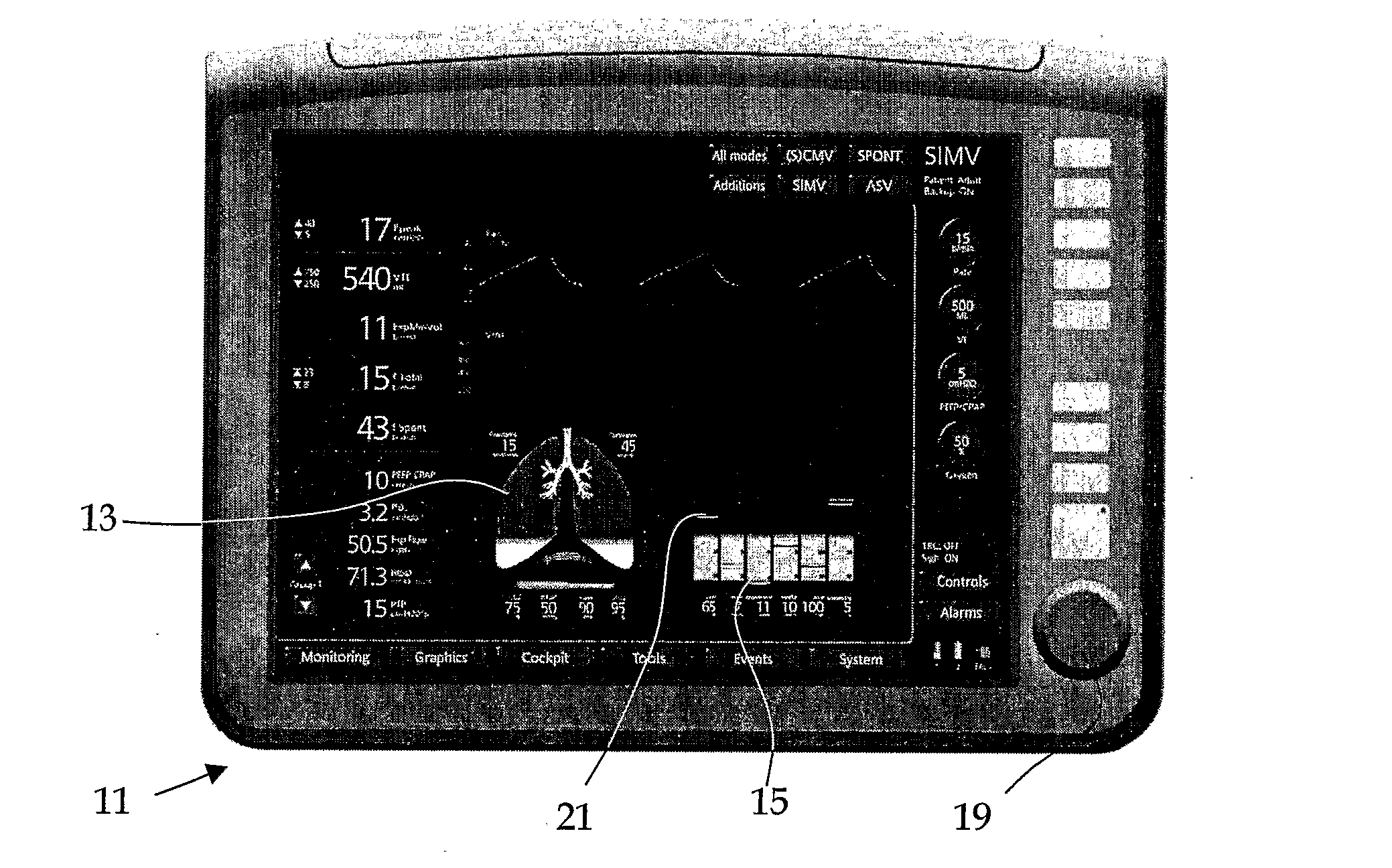
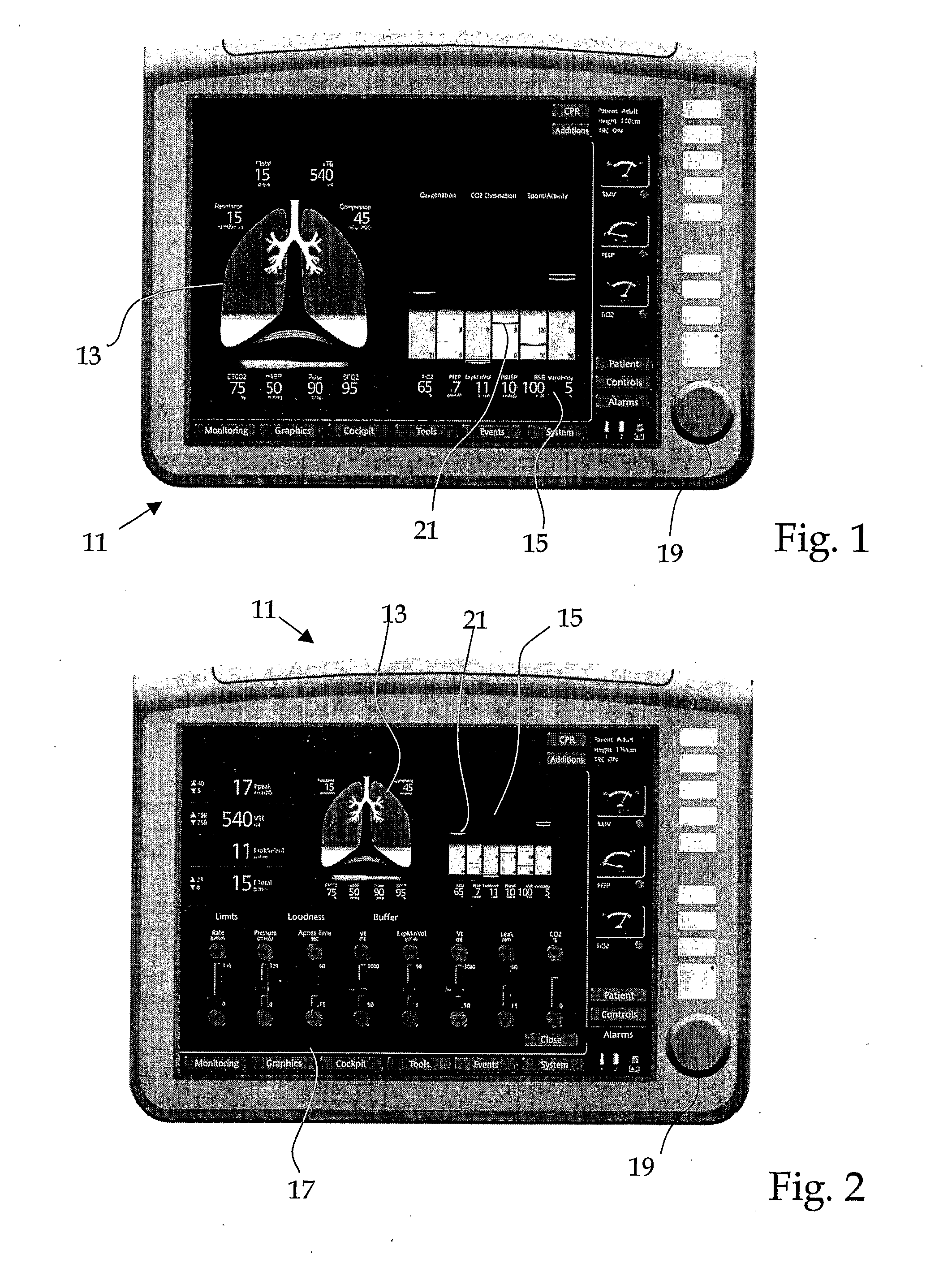
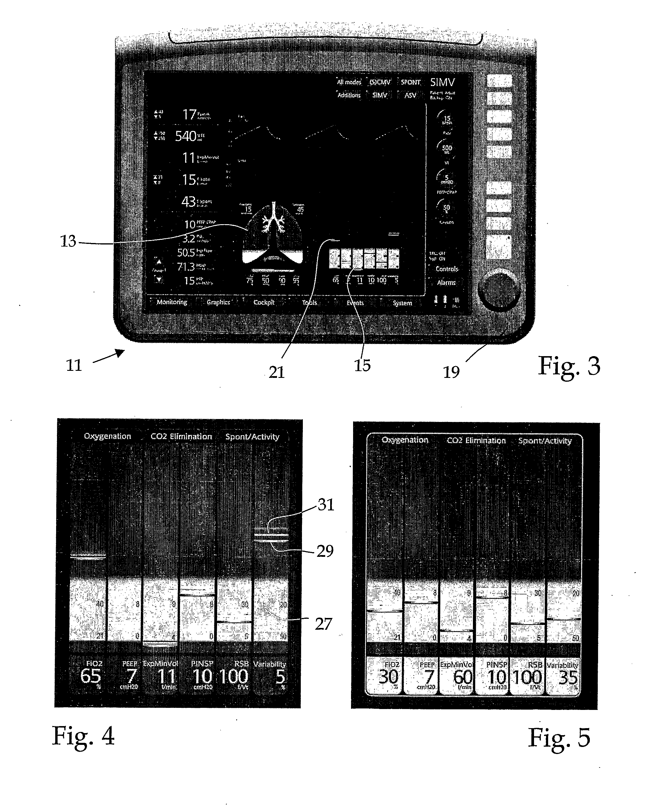
[0102]The screens 11 represented in the FIGS. 1 to 3 show representations generated with the method according to the invention. These representations comprise a first region 13 with patient parameters which contains an element reminiscent of an organ, and a region 15 with apparatus parameters which has a scale bar. The scales of this scale bar are different. Readings and apparatus settings are displayed on these scales with a positionally changing component 21. In FIG. 1, these two regions13 and 15 practically take up the complete screen. In FIG. 3 they are squeezed together in the upper half of the screen, since a window is opened around the lower region. Such a window may be selected and opened with the selection button 19, whereupon the representation according to FIG. 1 changes into a representation according to FIG. 2. In FIG. 3, the representation of both regions mentioned above only takes up a part region of the screen, whilst an upper part region pr...


PUM


Abstract

The invention relates to a method for acquiring several changing values and representing the acquired values on a monitoring screen, as well as to a device with a screen in order, on this, to represent changing values acquired during ventilation of the patient. The device comprises means for acquiring at least three changing values of different origin, and means for representing the values, which permit the acquired values to be qualitatively represented on the screen together in a single graphic element. This graphic element has a pictorial representation of a lung shape. The invention is characterised in that the means for representing the values are designed such that a volume change of the ventilated lung which is detected within each breath, is represented in an animated manner by way of size change of the lung shape corresponding to this corresponding volume change.

Description

TECHNICAL FIELD OF THE INVENTION[0001]The method relates to a method and to a device for acquiring several changing values which are be monitored during the ventilation of the patient, and representing the values on a screen.[0002]The mechanical ventilation of patients, as the case may be, is necessary to keep the patient alive. This is for example the case with an anesthetic, or also with diseases or after an accident. A mechanical ventilation may however also damage the lungs of the patient. It is therefore the object of any ventilation treatment, to carry it out as briefly as possible and with the lowest possible intensity. Patient parameters and apparatus settings are to be used for estimating the necessity of ventilation, as well as the necessary intensity of the ventilation.[0003]In order to adapt the ventilation where possible, to the requirements of the patient, a continuous, new assessment of the patient status and a likewise continuous, new assessment of the dependency of ...

Claims


Application Information

Patent Timeline
Patent Type & Authority Applications(United States)
IPC IPC(8): A61B5/08A61B5/01
CPCA61B5/087A61M16/0051A61M2016/0021A61M2205/584A61M2016/1025A61M2205/075A61M2205/502A61M2016/0036A61B5/0205A61B5/021A61B5/024A61B5/091A61B5/14542A61B5/743A61B5/08A61M16/00A61M16/021
Inventor BRUNNER, JOSEFWYSOCKI, MARCMETTIER, IVO
Owner HAMILTON MEDICAL AG
Why Eureka
Try Eureka