


Quick Research
Generate reliable direction feasibility study reports for your R&D in just a few steps.

Technical Q&A
Discover and master advanced knowledge NOW. Basics, ideas, possibilities, all at once.

Find Solutions
As an expert in R&D theories, this can generate solutions to your technical problems instantly.

Evaluate Feasibility
Analyze your overall solution with one click, know your potential R&D risks in advance.

Monitor Landscape
Get weekly tech updates, stay abreast of the latest tech innovations and key insights.
Abnormalities of Phosphatase 2A (PP2A) for Diagnosis and Treatment of Alzheimer's Disease
- Summary
- Abstract
- Description
- Claims
- Application Information
AI Technical Summary
Benefits of technology
Problems solved by technology
Method used
Image

Examples
example 1
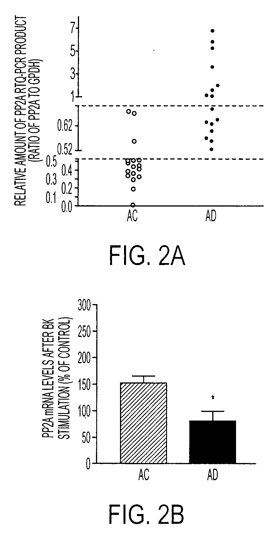
Changes in PP2A mRNA Levels in AD Cells
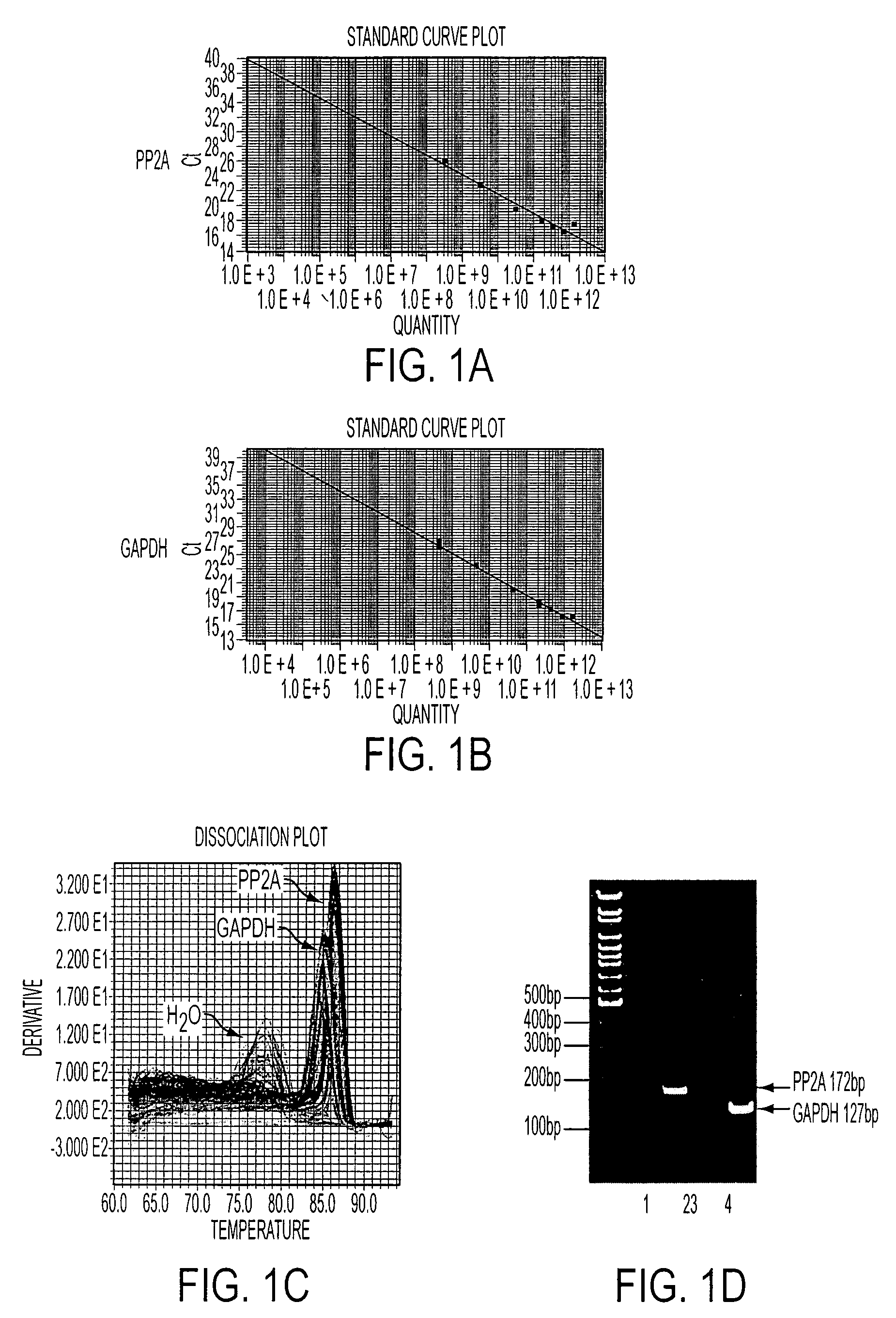
[0076]PP2A gene expression was quantified using RTQ-PCR, with GAPDH as a reference gene for normalization. As shown in FIGS. 1A and 1B, with real-time PCR, PP2A and GAPDH primers, respectively, produced a linear standard curve of the amplified sequence with a series of dilutions of the human fibroblast cDNA template run in duplicates. Specific melting temperatures (MT) were plotted by distinct dissociation curves (FIG. 2C) for PP2A, GAPDH, and water, demonstrating a high specificity of each PCR product. This specificity was confirmed by the result shown in FIG. 1D, in which the final PCR products for PP2A and GAPDH were run on a 10% TBE gel. A single band with the expected sequence size was revealed for each gene (lane 2 and 4), but it was not detected in the sample without adding reverse transcriptase during in vitro reverse transcription (lane 1 and 3). This indicates that the amplified PCR products for PP2A and GAPDH were not from the genomi...
example 2
Changes in PP2A Protein Levels and Enzymatic Activities in AD Cells
[0077]To determine whether changes in PP2A gene expression in AD cells were reflected in its protein expression and function, both PP2A protein levels and activity were compared between AC and AD cells. The amount of PP2A protein measured with Western blotting was significantly reduced in all AD cells compared to that in AC cells (P<0.01). This reduction of PP2A was not due to a lower amount of protein from AD cells that was loaded on SDS-gel, because levels of a reference protein annexin II from the same samples were not significantly different from those in AC cells (FIG. 3A). A consistent result of reduction of PP2A in AD cells was also produced when the PP2A-immunoreactive signals were normalized against the total protein loaded on the SDS-gel. In addition, PP2A activity was also markedly decreased in AD cells compared to AC cells (P<0.001) (FIG. 3B).
example 3
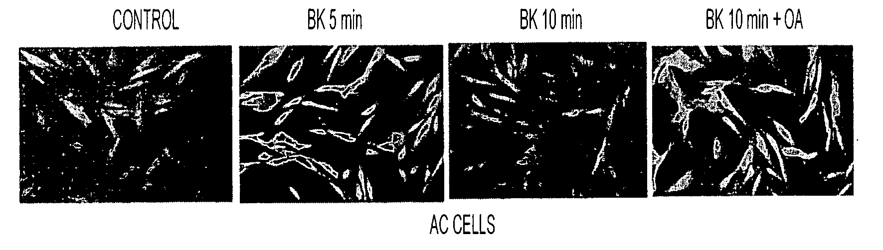
PP2A is Involved in Dephosphorylation of Erk1 / 2 after BK Stimulation
[0078]To test whether PP2A is involved in dephosphorylation of Erk1 / 2, AC cells from five different individuals were treated with a PP2A inhibitor, okadiac acid at a concentration only inhibiting PP2A (Nagao et al., 1995; Sheppeck et al., 1997; Fernandez et al., 2002). The Erk1 / 2 phosphorylation was determined on Western blots using specific antibodies for phospho- and regular Erk1 / 2. Erk1 / 2 phosphorylation was increased at about 5 min after BK stimulation, but it returned to the control level by about 10 min (FIG. 4) possibly due to a normal dephosphorylation mechanism in the cell. In the presence of about 10 nM OA, however, this Erk1 / 2 dephosphorylation was significantly inhibited (FIG. 4). A one-way ANOVA revealed significant treatment effects (P<0.001). These results indicate that PP2A is responsible for dephosphorylation of Erk1 / 2 after its BK-stimulated phosphorylation.
PUM
| Property | Measurement | Unit |
|---|---|---|
| Ratio | aaaaa | aaaaa |
| Gene expression profile | aaaaa | aaaaa |
Abstract
Description
Claims
Application Information
 - R&D Engineer
- R&D Manager
- IP Professional
- Industry Leading Data Capabilities
- Powerful AI technology
- Patent DNA Extraction
Browse by: Latest US Patents, China's latest patents, Technical Efficacy Thesaurus, Application Domain, Technology Topic, Popular Technical Reports.
© 2024 PatSnap. All rights reserved.Legal|Privacy policy|Modern Slavery Act Transparency Statement|Sitemap|About US| Contact US: help@patsnap.com