Fluorescent nanoscopy method

a fluorescence nanoscopy and nanoscopy technology, applied in the field of scientific research equipment and lens fluorescence microscopes, can solve the problem of hard to ensure the stability of molecular structures, and achieve the effects of improving resolution, ensuring stability of molecular structures, and low background noise level

Active Publication Date: 2009-02-19
SUPER RESOLUTION TECH
View PDF10 Cites 30 Cited by
  • Summary
  • Abstract
  • Description
  • Claims
  • Application Information

AI Technical Summary

Benefits of technology

The present invention relates to improving the resolution of biological imaging techniques like confocal microscopy. By combining the benefits of traditional optical microscopes with advanced technology, we have developed a method called“nanoscopy” that allows us to independently observe molecules within 10-22 nm of each other. This involves taking multiple pictures of lowly dyed objects and calculating the positions of individual molecules using computer analysis. Nanoscopy has applications in areas like medical diagnosis and materials science where understanding the structure of small particles is crucial.

Problems solved by technology

The technical problem addressed in this patent text is the limitations of conventional microscopes in identifying individual spots on an object's surface within a certain distance without using costly and complicated equipment. The current methods require either a minimum separation between the spots or multiple detection beams, resulting in slow scan speeds and limited accessibility to specific regions of the object.

Method used


Image

  • Fluorescent nanoscopy method
  • Fluorescent nanoscopy method
  • Fluorescent nanoscopy method

Examples


Embodiment Construction

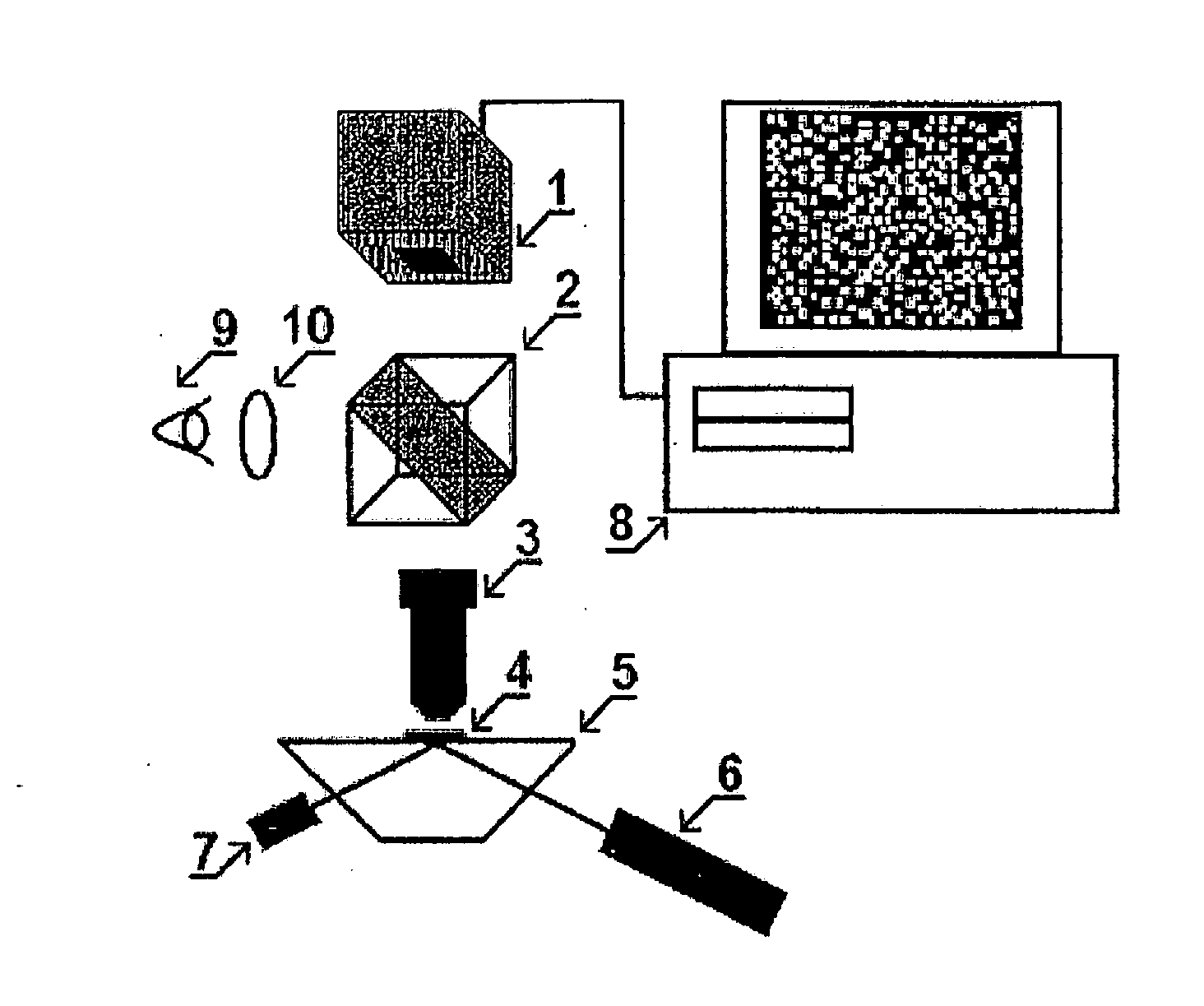
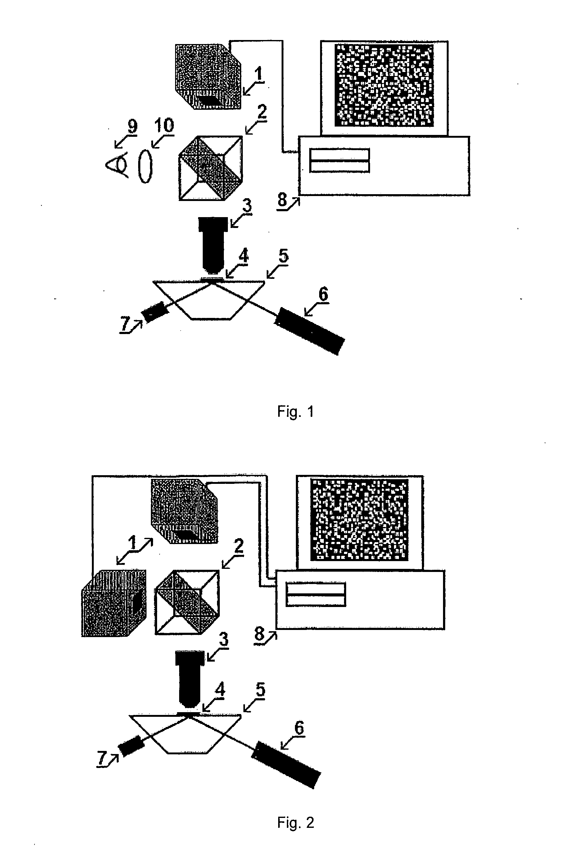
[0020]The fluorescent microscope-nanoscope, as it is shown on FIGS. 1 and 2, is equipped with: one (FIG. 1) and two (FIG. 2) monochrome video-cameras (1) with digital output and suppressing color-filters, located opposite to their sensors (CCD). These color-filters pass only fluorescent light to cameras. A microscope is also equipped with a light dividing removable prism (2); object lens (3) with zoom up to 100× and an aperture up to A=1.4. Object (4) is pressed down to glass object holder (5) which is beveled in the form of truncated prism edges. The device also has laser (6) with lens system for exciting fluorescence through prism planes; with a pulse UV-source with a lens system for photolysis of blocking fluorescence groups, present on dye molecules. Another important part is computer (8) with software for recording and working with digitized images. This software is also used for control of a power source, and providing energy for UV pulses and electrophoresis device. Eye lens ...


PUM


Abstract

An analysis of an object dyed with fluorescent coloring agents carried out with the aid of a fluorescent microscope which is modified for improved resolving power and called a nanoscope. The method is carried out with a microscope having an optical system for visualizing and projecting a sample image to a video camera which records and digitizes images of individual fluorescence molecules and nanoparticles at a low noise, a computer for recording and processing images, a sample holder arranged in front of an object lens, a fluorescent radiation exciting source and a set of replaceable suppression filters for separating the sample fluorescent light. Separately fluorescing visible molecules and nanoparticles are periodically formed in different object parts, the laser produces the oscillation thereof which is sufficient for recording the non-overlapping images of the molecules and nanoparticles and for decoloring already recorded fluorescent molecules, wherein tens of thousands of pictures of recorded individual molecule and nanoparticle images, in the form of stains having a diameter on the order of a fluorescent light wavelength multiplied by a microscope amplification, are processed by a computer for searching the coordinates of the stain centers and building the object image according to millions of calculated stain center co-ordinates corresponding to the co-ordinates of the individual fluorescent molecules and nanoparticles. With this invention it is possible to obtain a two-dimensional and a three-dimensional image with a resolving power better than 20 nm and to record a color image by dyeing proteins, nucleic acids and lipids with different coloring agents.

Description


Claims


Application Information

Patent Timeline
Owner SUPER RESOLUTION TECH
Why Eureka
Try Eureka