Method for rapid screening of mad cow disease and other transmissible spongiform encephalopathies
a spongiform encephalopathy and rapid screening technology, applied in the field of rapid screening of mad cow disease and other transmissible spongiform encephalopathies, can solve the problems of not being suited for in vivo testing, not optimal for rapid screening of large numbers of animals, and taking hours to compl
- Summary
- Abstract
- Description
- Claims
- Application Information
AI Technical Summary
Benefits of technology
Problems solved by technology
Method used
Image

Examples
example 1
CJD, Scrapie, and BSE Diagnosis Using Catheter Based OCT Probe to Visualize Vacuolar Appearance
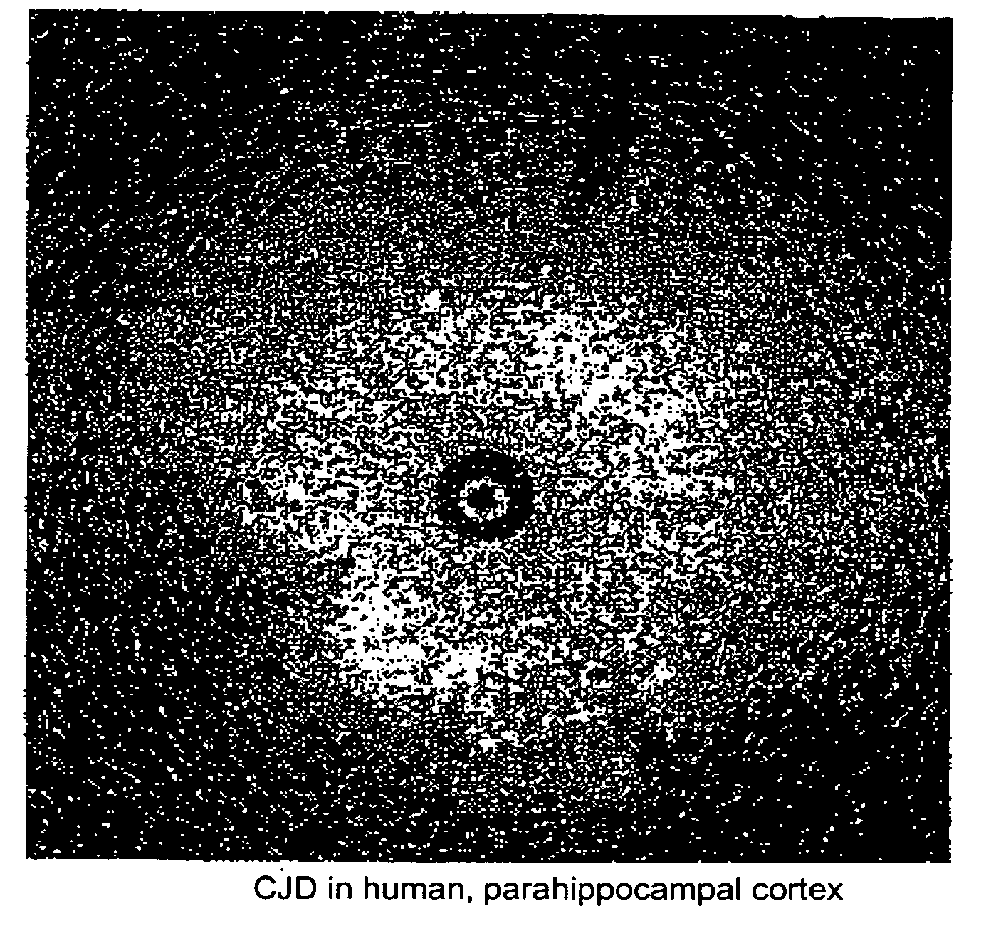
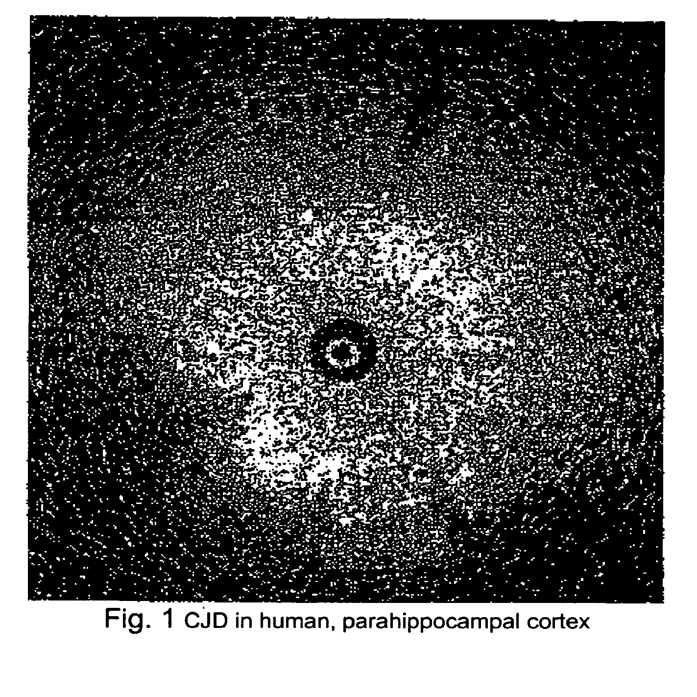
[0027]As illustrated in FIG. 1, brain tissue from a patient who died of CJD was imaged using a catheter based OCT probe manufactured by LightLab Imaging (of Westford, Mass.). Large numbers of vacuoles of different diameters were observed. The high degree of back scattering by the vacuoles suggests that they are not simple vacuoles filled with CSF-like fluid. Vacuoles having the observed OCT appearance shown in FIG. 1 have not been observed in human brain stored in the same manner.
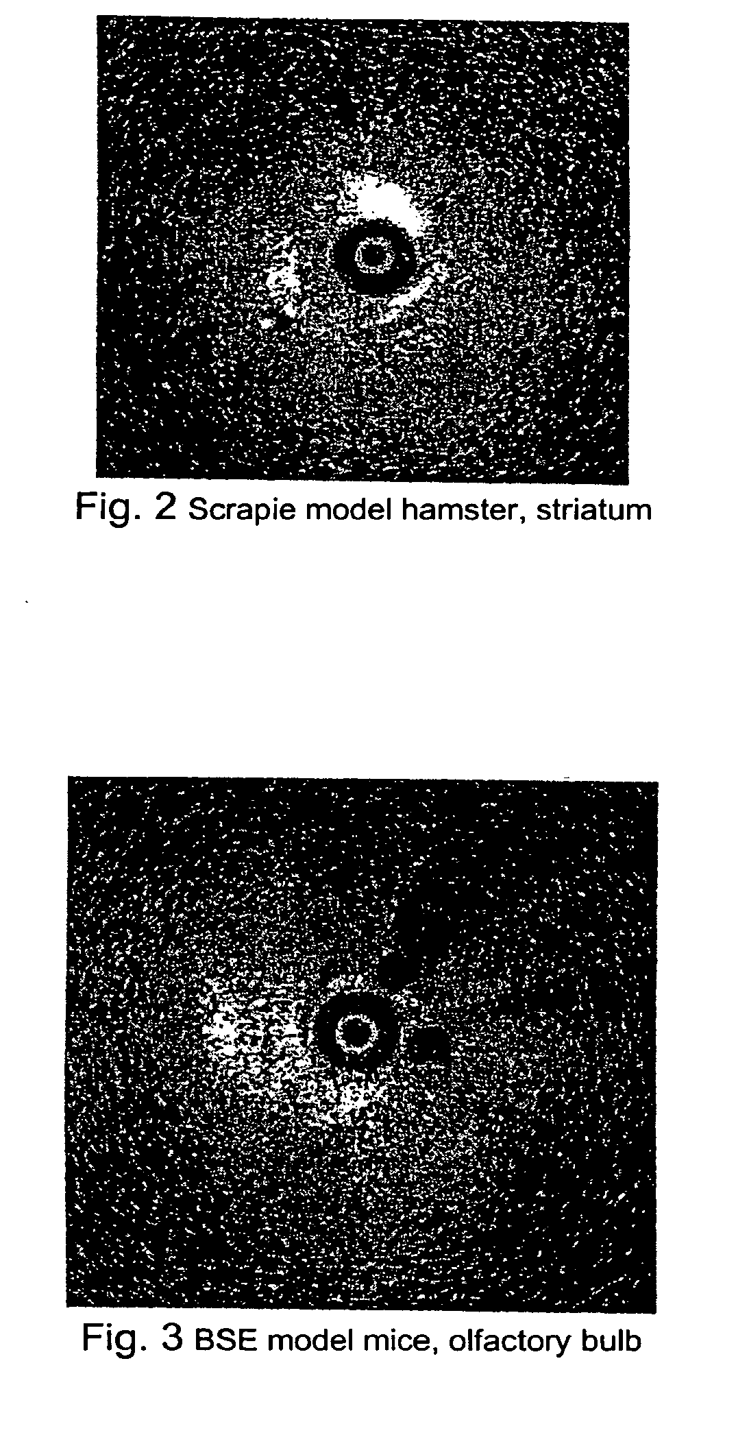
[0028]As illustrated in FIG. 2, a hamster infected with scrapie was sacrificed shortly before OCT imaging. Highly reflective vacuoles similar to that observe in CJD brain were observed in the striatum and possibly in the cortex.
[0029]As illustrated in FIG. 3, OCT was performed in a mouse brain infected with BSE. Large vacuoles were identified in the olfactory bulb.
example 2
Methods for Screening Tissue of Slaughtered Animals
Imaging Using a Needle-Type Probe
[0030]A catheter-based OCT probe packaged within a rigid cannula (needle-type probe) is inserted into an exposed tissue (i.e. brain, spinal cord, etc) of a slaughtered animal. The approach is used when tissue deep in the brain is desired for sampling and / or testing. A needle type probe may also be inserted directly through thin regions of the skull (i.e. through the roof of orbit below the eye brow to sample the frontal cortex). A radial scan may be performed to image the brain as illustrated in the proceeding figures. The probe will be advanced to sample a volume of tissue. The data may be interpreted by the operator in real time or may be stored for off-line processing. Software may be developed to automatically identify, measure, and count the number of vacuole per volume of tissue sampled. The index of refraction of the vacuole may also be determined based on the amplitude of reflected light. The...
example 3
Methods for Screening Tissue of a Slaughtered Animal
Contact but Non-Penetrating Imaging
[0031]A clear disposable window may be placed against the tissue to separate the OCT probe from the brain. These probes may or may not need to be catheter based. Catheter-based probes may have a linear scanning movement, similar to the ‘push-pull’ design of LightLab Imaging and probes currently designed for GI endoscopy and dermatology. Non-catheter-based probes may use designs similar to those used for OCT opthalmoscope and OCT microscope. This method is best suited for pathology that is located at a relative short distance from the surface of the tissue. Most spongiform lesions in the cortex are within the detection distance from the surface of the cortex. It is also possible to cut the sample so that pathology anywhere within the brain may be detected. In such case, the tissue would need to be handled but still would not need to be extensively prepared as for conventional histology.
PUM
 Abstract
Description
Claims
Application Information
 - R&D
- Intellectual Property
- Life Sciences
- Materials
- Tech Scout
- Unparalleled Data Quality
- Higher Quality Content
- 60% Fewer Hallucinations
Browse by: Latest US Patents, China's latest patents, Technical Efficacy Thesaurus, Application Domain, Technology Topic, Popular Technical Reports.
© 2025 PatSnap. All rights reserved.Legal|Privacy policy|Modern Slavery Act Transparency Statement|Sitemap|About US| Contact US: help@patsnap.com