Flexible visually directed medical intubation instrument and method

a visual directed, flexible technology, applied in the field of medical instruments, can solve the problems of urethral obstruction, difficult placement of more than forty percent of male urinary catheters, burden on the delivery of effective care through the healthcare system, etc., and achieve the effect of reducing the cost of the instrumen

Inactive Publication Date: 2009-12-24
PERCUVISION
View PDF79 Cites 179 Cited by
  • Summary
  • Abstract
  • Description
  • Claims
  • Application Information

AI Technical Summary

Benefits of technology

[0006]Yet another object is to provide a surgical intubation instrument for providing visually directed placement into the body of the patient that makes possible a dramatic reduction in the cost of the instrument.

Problems solved by technology

While most placements proceed without problems, typically more than forty percent of male urinary catheter placements are difficult because of the problematic normal anatomy of the male lower urinary tract such as the external sphincter, the S-curve of the bulbous urethra and angulated prostatic urethra and / or pathologic conditions, such as urethral stricture disease, stones, trauma, tumors, enlarged prostate, iatrogenic false passages, and / or congenital disorders causing a substantial burden on the delivery of effective care through the healthcare system.
The most common problem is tetany, a spasm of the external urinary sphincter or stricture of the urethra.
Stones, and even clots descending from the bladder, also constitute urethral obstructions.
These costs to the healthcare system, hospitals, clinics and doctors' offices are substantial.
In addition, the delay in servicing urological catheter patients in a timely manner constitutes poor medical efficiency, delivery, and control.
When difficulty is encountered, the resulting frustration among healthcare professionals, especially nurses, physician extenders and physician assistants, creates a very real feeling of ineffectiveness on the part of these healthcare workers, to say nothing of the dissatisfaction on the part of the patients caused by the delay and added discomfort.
Difficult catheterizations can also be a source of urinary tract infection.
While the dollar cost to the healthcare system is not the only concern, such elements as added labor and material costs, time delays for patient rectification, excess space and equipment required, catheter kit value, nurse technician and physician costs constitute an expense to the healthcare system of surprising proportions.
Of these, conservatively about 40% are difficult which means that they require multiple advances and pull-backs of the urinary catheter to negotiate the urethra, multiple catheters on the same patient, several staff workers attending to the same patient, or special instrumentation such as filoforms / followers, cystoscope or radiologic services.
Two prior U.S. patents by the present inventor; U.S. Pat. Nos. 6,599,237 and 6,994,667 are directed to some of these problems and, while they provide excellent results, they are not ideal in all applications, have some limitations in specific areas of use, and cannot therefore be considered completely versatile with respect to their application in certain surgical specialties.
Another important consideration is the high cost of surgical instruments, which may be from several hundred to several thousand dollars.
Other instruments may be suited for urological use but not be suited for use in gastroenterology.
Certain intubation devices such as the Councill catheter are only capable of a blind insertion and must rely on a guide wire to navigate to the bladder.
Consequently, if the Councill catheter encounters resistance during insertion, there is no way to know its cause.
Any obstructions, anomalous conditions, or anatomical idiosyncrasies can interfere with successful insertion of the tube.
Moreover, no provision is made for sensing conditions at or near the distal tip of the intubation instrument with traditional analog sensors and / or actuators or smart digital sensors or actuators.

Method used


Image

  • Flexible visually directed medical intubation instrument and method
  • Flexible visually directed medical intubation instrument and method
  • Flexible visually directed medical intubation instrument and method

Examples


Embodiment Construction

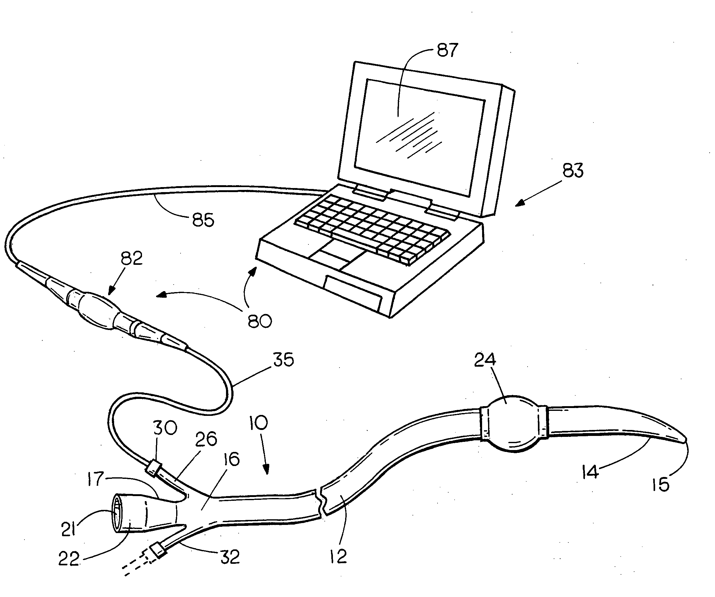
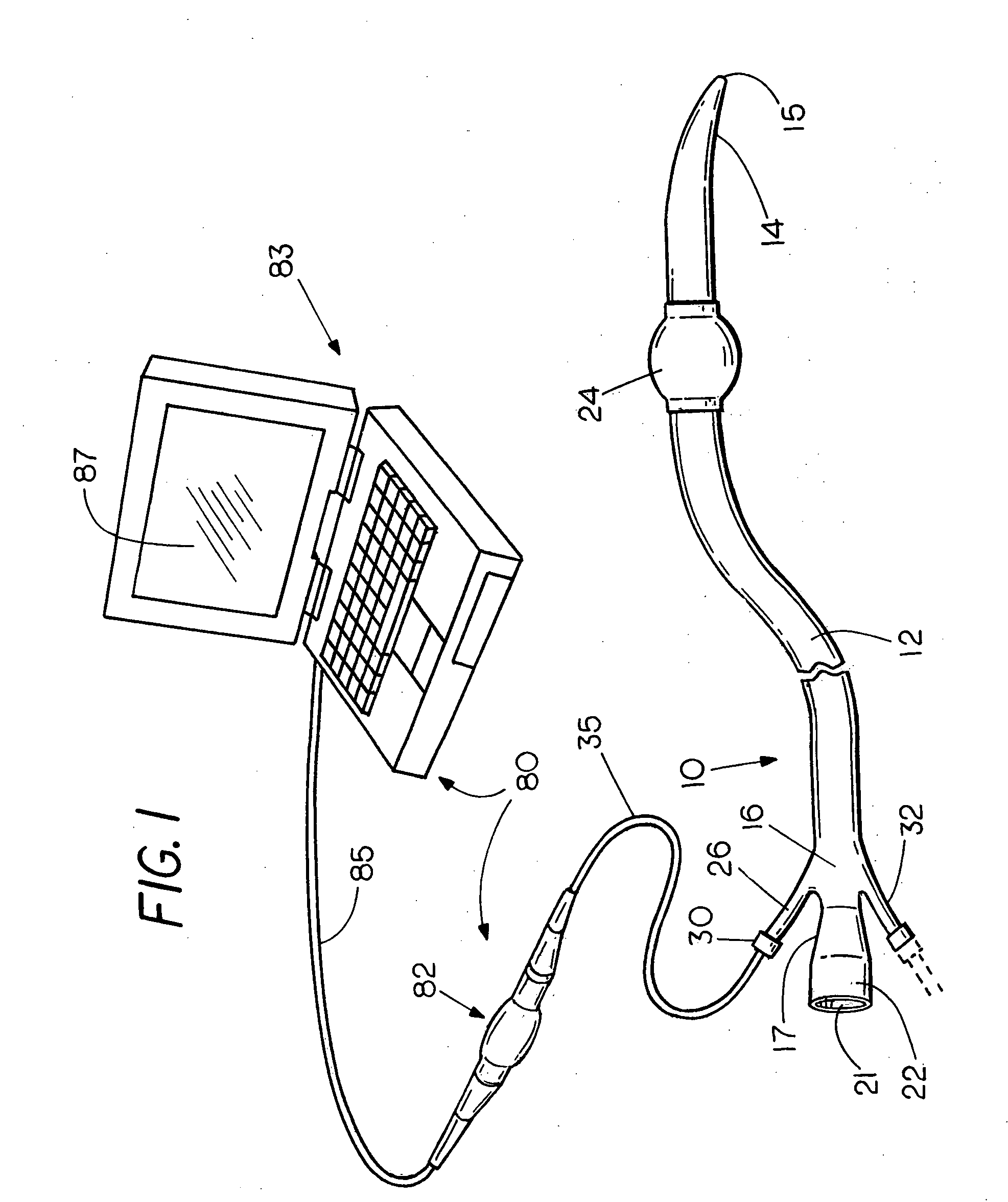
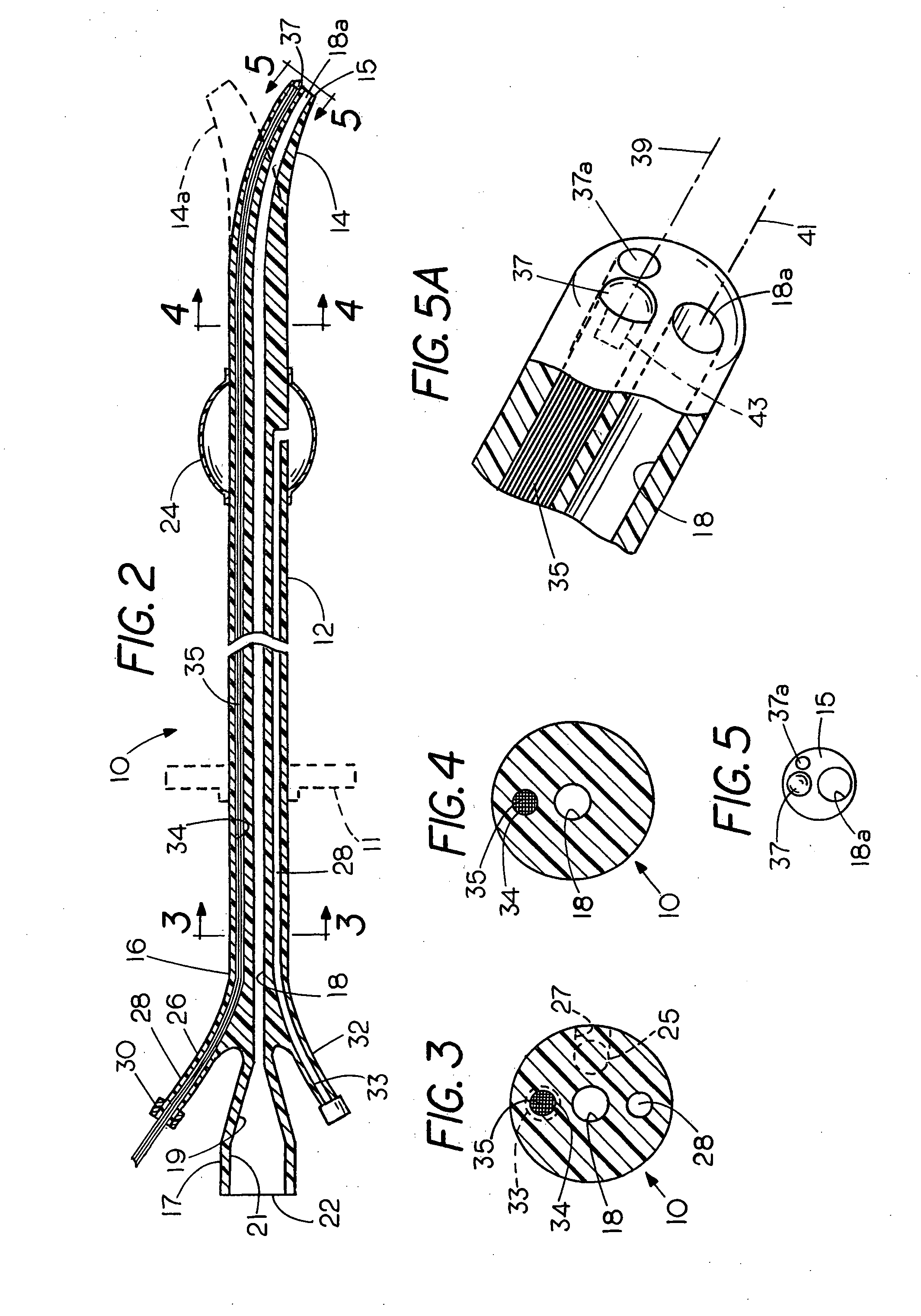
[0029]Refer now to the Figures wherein the same numerals refer to corresponding parts in the several views. The invention will be described by way of example with reference to FIGS. 1-7 and 14 which illustrate a visually directed intubation instrument in accordance with the invention that can be placed into the body of the patient under direct and continuous visual control in any of a variety of different surgical specialties. The invention is especially versatile and can be dimensioned and configured for use in urology, in gastroenterology, and in other surgical fields. The embodiment of FIGS. 1-7 and 14, illustrate the versatility of the invention since it can be employed as a drain or for exploratory purposes as well as a working channel to be used during a surgical operation or even in the field of gastroenterology as a feeding tube.

[0030]The instrument 10 comprises a flexible catheter 12 formed from natural or synthetic rubber or from a flexible biocompatible polymer of any sui...


PUM


Abstract

A flexible medical intubation instrument provided for placement into an animal or human patient comprises a catheter with at least a pair of longitudinally extending lumens or channels including a sensor and/or actuator channel and a working channel. In the sensor/actuator channel is provided a fixed or slideably removable sensor cable having a sensor for sensing a characteristic or condition including any of the following: a visual sensor for optical viewing, a chemical sensor, a pH sensor, a pressure sensor, an infection sensor, an audio sensor, or a temperature sensor. The sensors are coupled by the sensor/actuator cable through light transmission, electric current, or radio transmission to a viewing instrument or other output device such as a meter or video screen for displaying the condition that is sensed within the body of the patient while the flexibility of the composite structure comprising the catheter and cable enable the entire instrument to flex laterally as it moves through curved passages or around obstructions during insertion or removal. While making observations through the sensor channel, the working channel simultaneously functions as a drain or an irrigation duct, a feeding tube, or to provide a passage for the insertion of one or a succession of surgical devices such that the catheter serves as a protective artificial tract or liner as surgical devices are inserted and removed through it in succession so as to minimize tissue trauma, infection, and pain experienced by the patient. The instrument can be used in urology, as well as a visually directed nasogastric tube, as a visually directed external gastrostomy tube, or as a visually directed internal gastric tube or percutaneous endoscopic gastrostomy tube and in other applications.

Description

FIELD OF THE INVENTION[0001]This invention relates to medical instrumentation and more particularly to a method and apparatus for facilitating intubation of an animal or human patient.BACKGROUND OF THE INVENTION[0002]In many medical procedures it is often necessary to place an instrument into the body of the patient for drainage, for viewing a part of the body, or for performing a surgical operation such as the endoscopic removal of a tumor, to take a biopsy, or for feeding the patient. The invention has general application in medicine including the field of urology as well as in the field of gastroenterology and in other medical and surgical specialties. The placement of a catheter in the urethra for the purpose of draining urine or for diagnostic purposes, for example, is one of the most common urological procedures for draining urine or fluid to determine the amount of urine present, to diagnose problems, or to maintain anatomic continuity. This procedure is commonly performed by...

Claims


Application Information

Patent Timeline
Patent Type & Authority Applications(United States)
IPC IPC(8): A61B1/04
CPCA61B1/00165A61B1/012A61B1/07A61J15/0026A61B5/036A61M25/0105A61B5/0084
Inventor SINGH, ERROL O.
Owner PERCUVISION
Why Eureka
Try Eureka