
Closure Device for Skull Plates and Related Method Thereof

a technology of closure device and skull plate, which is applied in the field of medical implants, can solve the problems of not being able to achieve rapid and convenient means or methods for neurosurgeons, and achieve the effect of convenient and effective repositioning of the bone flap

Active Publication Date: 2010-02-18
UNIV OF VIRGINIA ALUMNI PATENTS FOUND
View PDF32 Cites 6 Cited by
  • Summary
  • Abstract
  • Description
  • Claims
  • Application Information

AI Technical Summary

Benefits of technology

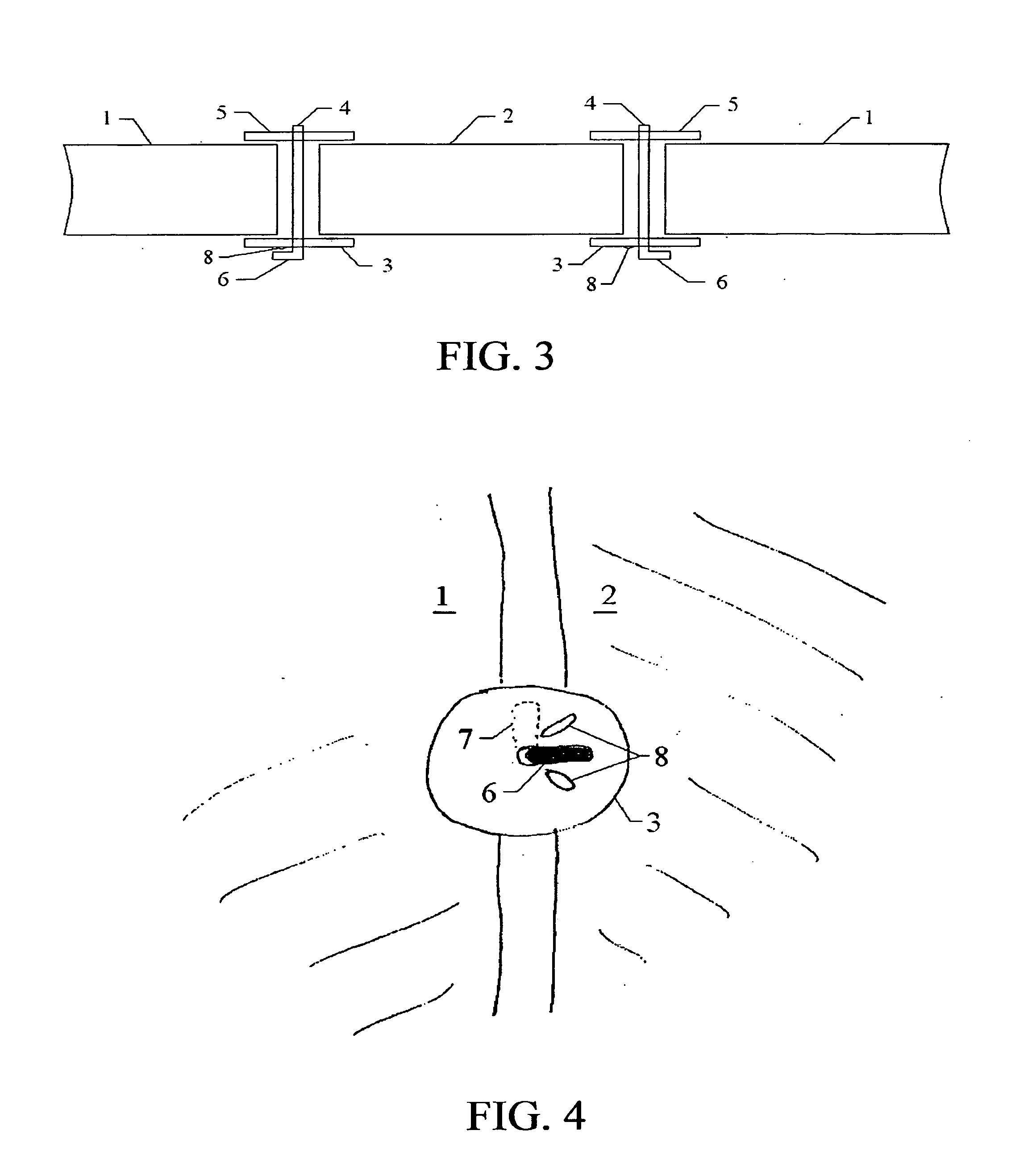
[0005]To overcome these and other limitations, an aspect of various embodiments of the present invention provides a method, device, and means for obtaining fixation of a bone flap within an aperture in the skull that allows for easy and effective repositioning of the bone flap. An approach disclosed herein uses sets of capture plates to laterally position the bone flap within the aperture, with a shaft intersecting each opposing pair of capture plates. The sets of capture plates may be positioned circumferentially around the outer perimeter of the bone flap, at locations deemed most appropriate by the neurosurgeon or practitioner. Flexibility intrinsic to the upper capture plate allows the interconnection shaft to be extended beyond the distal surface of the lower plate, and thus rotated into a detenting lock position or, alternatively, into a detenting unlock position for the loosening or withdrawal of the shaft and repositioning of the bone flap.
[0007]An aspect of an embodiment of the present invention provides a device for fixation of an excised piece of hard or firm tissue within an aperture in a larger hard or firm tissue region of a patient. The device comprising: a lower plate to be positioned below the excised piece of hard or firm tissue; a shaft having a distal end and a proximal end; an arm located at the distal end of the shaft, wherein the arm extends transversely from the shaft; and an aperture in the lower plate, the aperture adapted to allow passage of the shaft through the lower plate in an inward direction and when the shaft is rotated the arm rotates to a position preventing passage of the shaft through the lower plate in an outward direction.
[0009]An aspect of an embodiment of the present invention provides a method for fixating an excised piece of hard or firm tissue within an aperture in a larger hard or firm tissue region of a patient. The method comprising: positioning a lower plate below the excised piece of hard or firm tissue, the lower plate having an aperture therein; providing a shaft having a distal end and a proximal end, the shaft having an arm that is located at the distal end of the shaft, wherein the arm extends transversely from the shaft; passing the shaft through the lower plate in an inward direction; rotating the shaft; and preventing passage of the shaft through the lower plate in an outward direction.

Problems solved by technology

A significant limitation of these methods is that they do not allow for a rapid and convenient means or method for the neurosurgeon to de-couple the capture plates, thus enabling either additional access to the brain tissues or re-adjustment of the bone flap position.

Method used


Image

  • Closure Device for Skull Plates and Related Method Thereof
  • Closure Device for Skull Plates and Related Method Thereof
  • Closure Device for Skull Plates and Related Method Thereof

Examples


Embodiment Construction

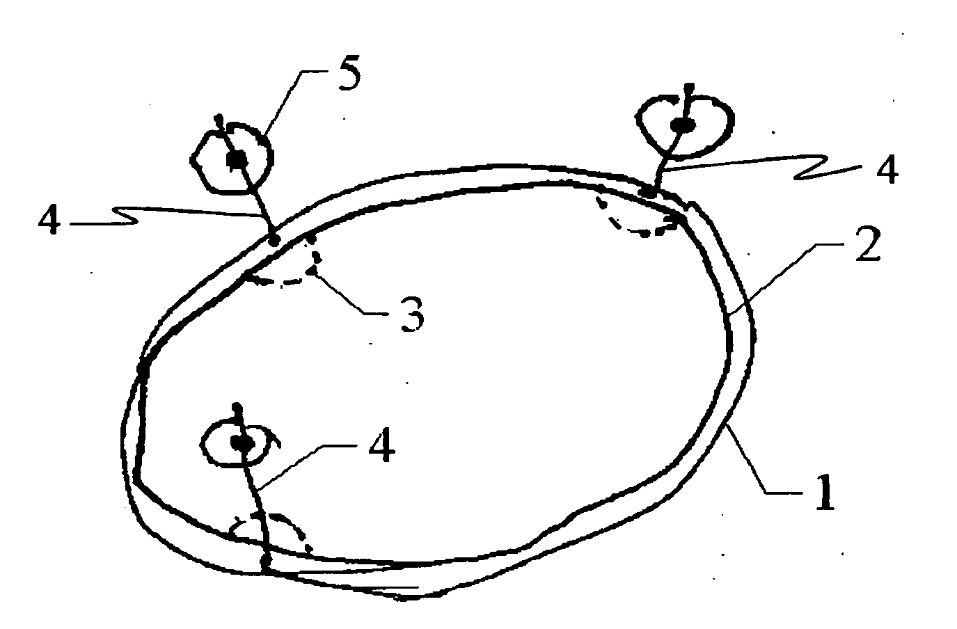
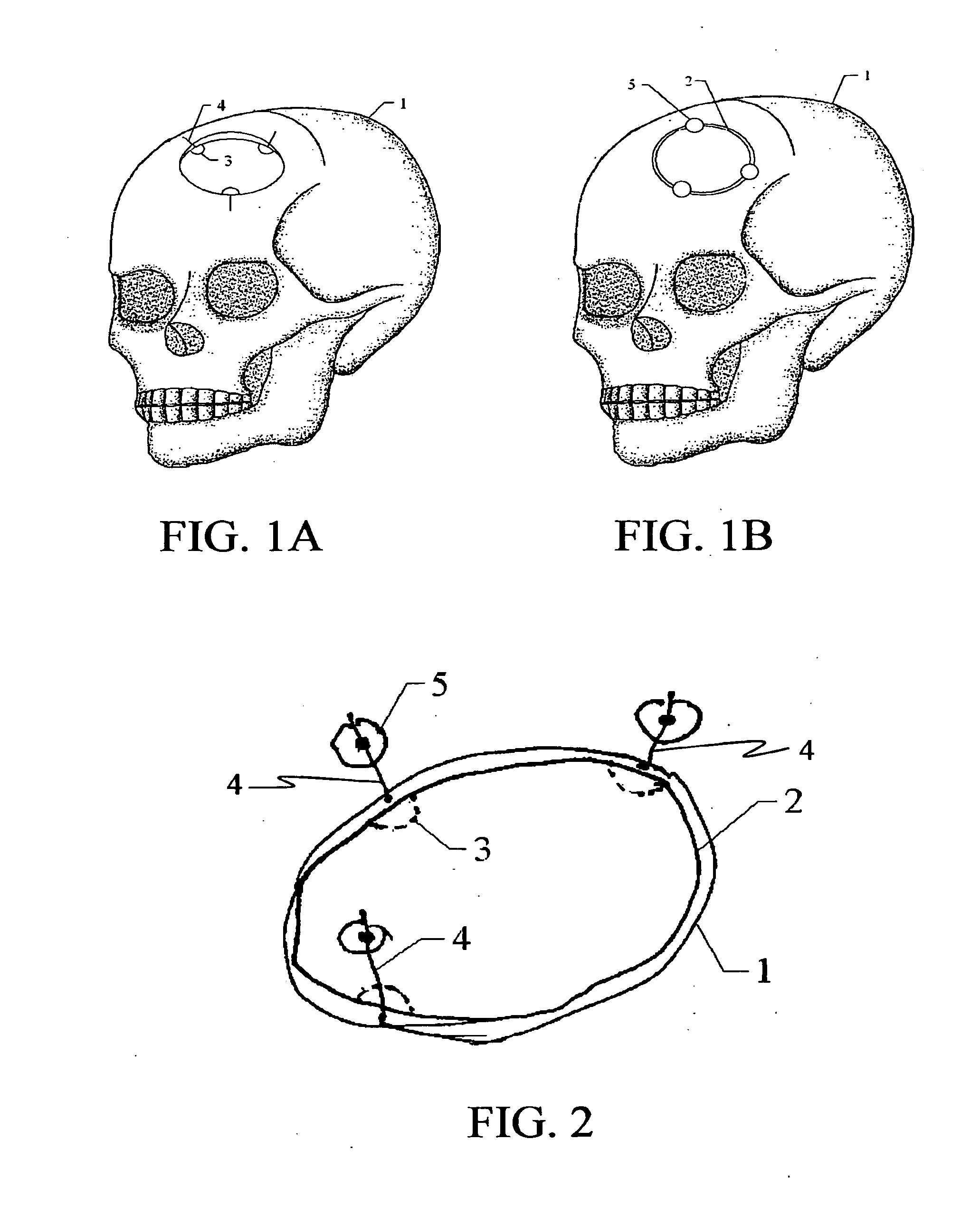
[0025]FIG. 1(A) shows a schematic view of a section of the skull 1 from which a circular bone flap 2 has been cut out to / y be any variety of shapes, but a circular shape is shown in order to simplify the illustration of the concept. An embodiment of the invention, consisting of a lower plate 3 and a shaft 4, is placed at various locations within the aperture in the skull 1. The shaft 4 is shown as inserted before the bone flap 2 is inserted into the aperture in the skull 1, but can also be inserted after the bone flap 2 is inserted. FIG. 1B shows a schematic view of the bone flap 2 positioned in the desired location within the aperture in the skull 1. An upper plate 5 is positioned above the bone flap 2 and in communication with the shaft 4 in order to secure the bone flap 2 within the skull 1. The upper plate 5 may be attached to the shaft 4 before or after passage of the shaft 4 through the lower plate 3. The upper plate 5 may be retained against the bone flap 2 and / or the skull 1...


PUM


Abstract

A method and means of cranial bone flap fixation that provides, among other things, the re-opening, resetting and / or repositioning of the bone flap during neurosurgical procedures. The instrumentation central to this method and means is MR and CT-visible to aid in imaging-based localization of it. The method and means can also be used in other types of medical procedures where certain kinds of hard or firm tissue fixation is desirable according to the method of the invention.

Description

CROSS-REFERENCE TO RELATED APPLICATIONS[0001]The present application claims benefit of priority from U.S. Provisional Application Ser. No. 60 / 816,716 filed Jun. 27, 2006, entitled “Closure Device for Skull Plates and Related Method Thereof,” wherein the disclosure is hereby incorporated by reference herein in its entirety.FIELD OF THE INVENTION[0002]This invention relates generally to medical implants and methods used to fix excised hard tissue secondary to the repair of an injury or the completion of a surgical procedure. More specifically, this invention may be utilized for fixing cranial bone flaps in place following neurosurgical procedures.BACKGROUND OF THE INVENTION[0003]Bone flaps are typically reattached into an opening in the skull following a craniotomy or other type of neurosurgical operation using clips, fasteners, and various other types of interconnection instrumentation that penetrate the bone, as well as posts that couple to capture plates that can laterally position...

Claims


Application Information

Patent Timeline
Patent Type & Authority Applications(United States)
IPC IPC(8): A61B17/80
CPCA61B17/688A61F2250/0098A61F2/2875A61F2002/3008A61F2002/30113A61F2002/30153A61F2002/30154A61F2002/30156A61F2002/30225A61F2002/30329A61F2002/30438A61F2002/30441A61F2002/30448A61F2002/30472A61F2002/3052A61F2220/0025A61F2220/0041A61F2220/005A61F2230/0006A61F2230/0019A61F2230/0021A61F2230/0023A61F2230/0069A61B19/54A61B90/39A61F2002/30433
Inventor BROADDUS, WILLIAM C.CHEN, ZHI-JIANGILLIES, GEORGE T.
Owner UNIV OF VIRGINIA ALUMNI PATENTS FOUND
Learn More