
Urological medical device and method for analyzing urethral properties

a medical device and urethral technology, applied in the field of urological medical devices, can solve the problems of inaccurate measurement of vlpp, lack of prior art devices, and lack of clear cut anatomic landmarks

Inactive Publication Date: 2010-03-18
BLAIVAS JERRY G
View PDF16 Cites 15 Cited by
  • Summary
  • Abstract
  • Description
  • Claims
  • Application Information

AI Technical Summary

Benefits of technology

[0063]In an exemplary embodiment, the urological medical device includes a control unit in communication with the pressure sensors and one or more sensors on the elongate body adapted to sense fluid. The elongate body is placed in the urethra and the control unit is adapted to at least one of (i) compute a vesical leak point pressure (VLPP), (ii) determine a point in the urethra where urethral pressure (Pure) becomes greater than vesical pressure (Pves), (iii) compute bladder neck descent during incontinence, (iv) compute a resting urethral angle, (v) compute a urethral angle during incontinence, (vi) compute a compliance of the urethra during incontinence, (vii) track movement of various points along a length of the elongate body during incontinence and determine which point moves the most, (viii) determine an anatomic location of the urethral meatus, and (ix) determine an anatomic location of a proximal extent of the sphincter.
[0064]Reference throughout this specification to “an embod

Problems solved by technology

However, there are no clear cut anatomic landmarks, either macroscopically or microscopically that delineate the sphincter.
However, merely knowing the diagnosis only permits a general approach to treatment.
This may result in inaccurate measurement of VLPP.
The prior art devices are also lacking in that they do not measure urethral compliance in a physiologic manner and do not assess abnormalities of the mucosal seal.
Further, neither the Regnier et al. nor the Lose probe are capable of (i) measuring the relevant physiologic properties at more than one place in the urethra at a time or (ii) measuring changes due to urine entering the urethra.
Existing devices are also deficient in that they do not properly assess urethral mobility.
Abnormalities of urethral support have been attributed to weakness of the pelvic floor muscles and fascia, primarily subsequent to pregnancy and childbirth.
Thirdly, there may be no rotation at all, but during increased abdominal pressure, the urethra may widen and shorten.
The latter two conditions result in incontinence.
The classic methods of assessing urethral mobility are the Q-tip angle and the chain cystogram, but both techniques only measure urethral angle and descent and do not assess the relationship between the two parameters, i.e., the urethral angle and concomitant urethral pressure at which urine leakage occurs, nor do they assess urethral shape or compliance, or define the axis around which the urethra rotates.

Method used


Image

  • Urological medical device and method for analyzing urethral properties
  • Urological medical device and method for analyzing urethral properties
  • Urological medical device and method for analyzing urethral properties

Examples


Embodiment Construction

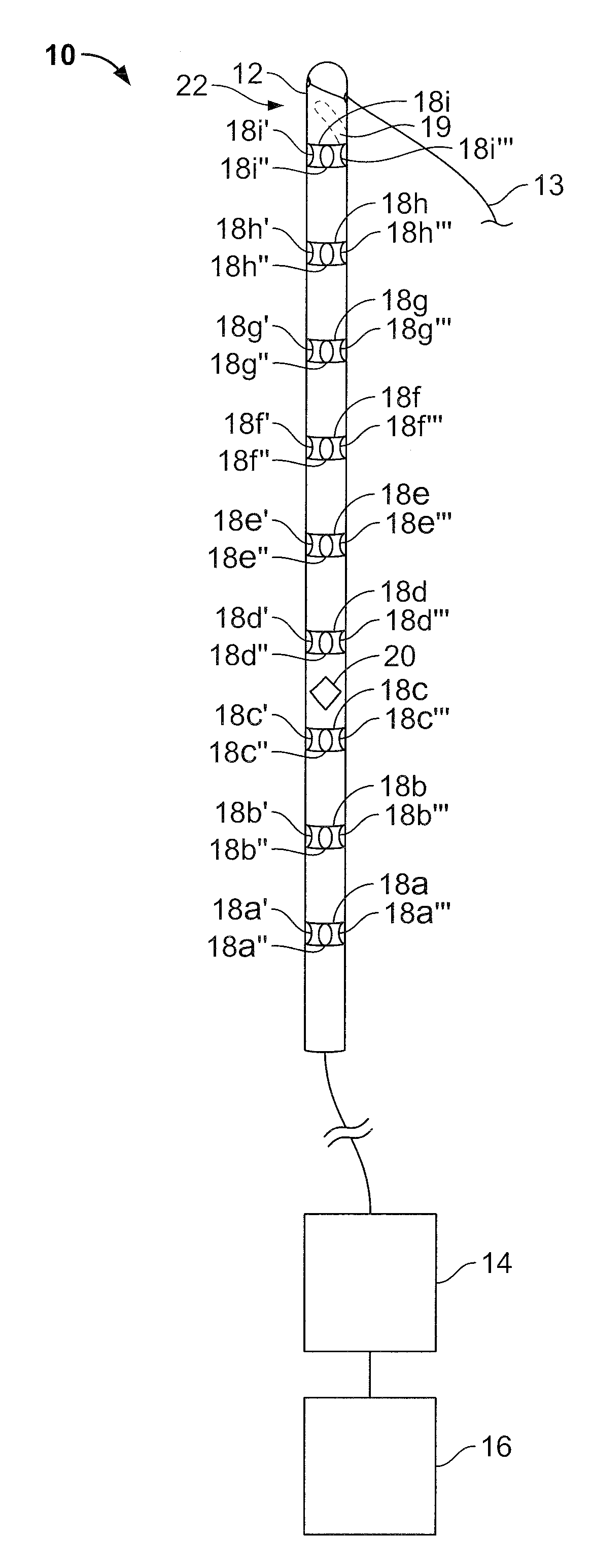
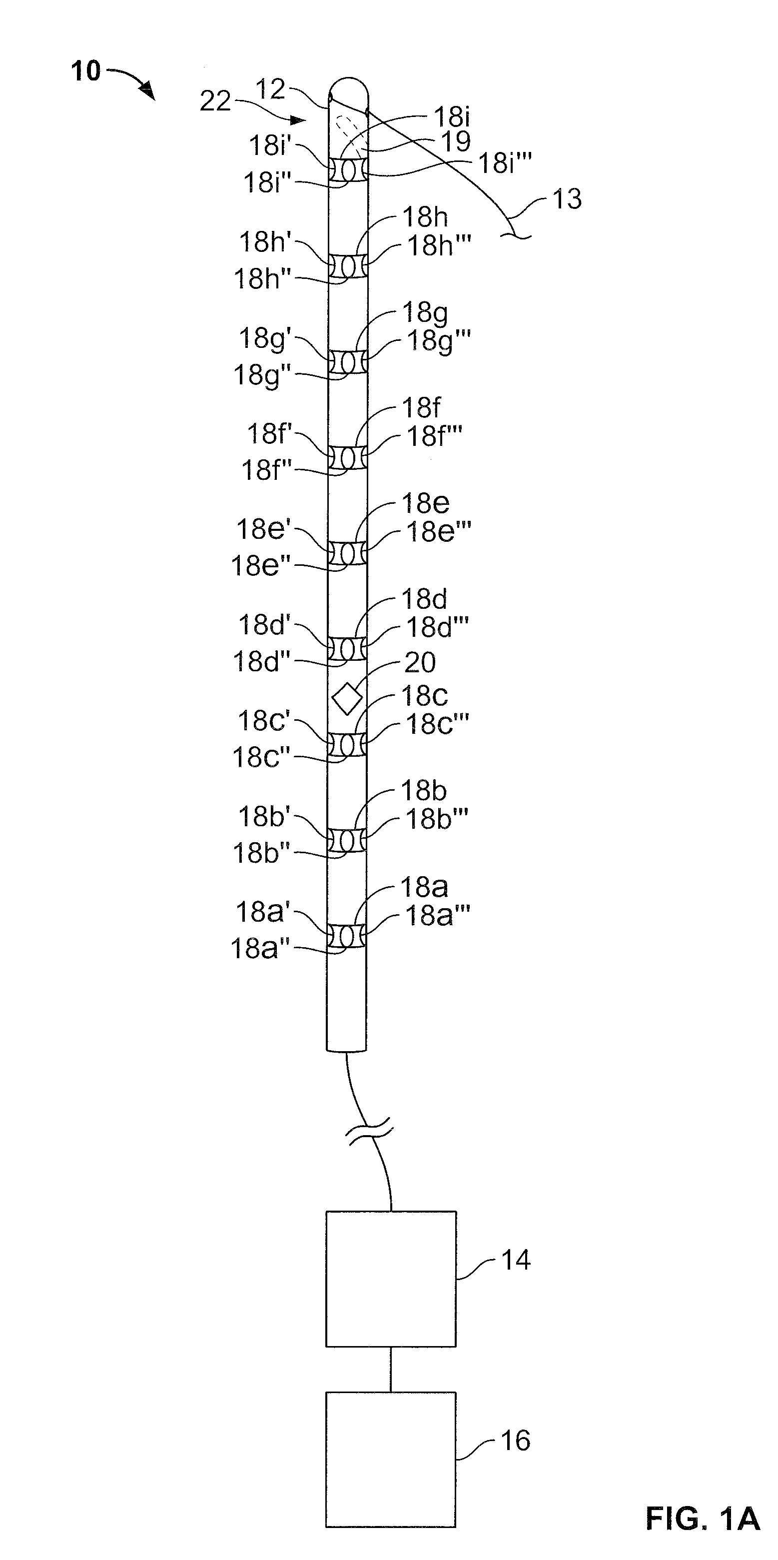
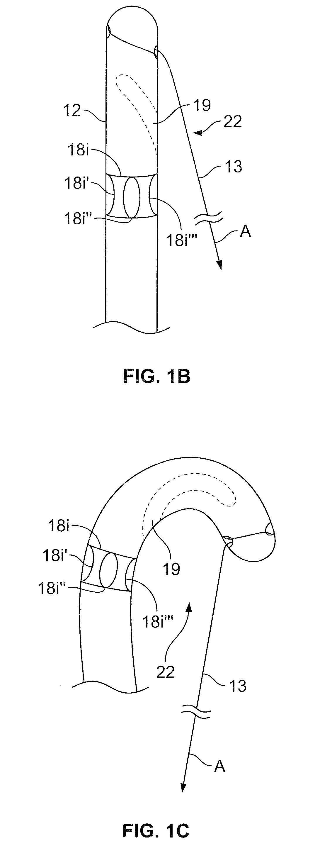
[0072]FIG. 1 is a perspective view of an urological medical device according to an exemplary embodiment of the present invention. The device includes an elongate body 10 adapted to be inserted into the urethra of a patient, proximal end 12 first. The elongate body 10 communicates with a control unit 14, which in turn communicates with a monitor display 16, which can be separate from or integrated with the control unit 14. As illustrated the elongate body 10, control unit 14, and display 16 have a hardwire and / or physical connection but they may also communicate wirelessly.

[0073]The elongate body 10 can be inserted into a patient, for example, using a stiff insertion rod. A proximal end of the rod is disposed in a lumen 19 (shown in dashed lines) and advanced into the urethra with the elongate body 10 until the proximal end 12 lies in the bladder of the patient. Other methods for insertion may be used as well, including the use of an insertion sheath. The elongate body 10 may also be...


PUM


Abstract

A highly flexible urological medical device for analyzing urethral properties and for optimally placing a periurethral injection and / or a support element, such as suburethral sling and / or a suspension suture, to treat urinary incontinence.

Description

CROSS-REFERENCE TO RELATED APPLICATIONS[0001]Not Applicable.STATEMENT REGARDING FEDERALLY SPONSORED RESEARCH OR DEVELOPMENT[0002]Not Applicable.THE NAMES OF THE PARTIES TO A JOINT RESEARCH AGREEMENT[0003]Not Applicable.INCORPORATION-BY-REFERENCE OF MATERIAL SUBMITTED ON COMPACT DISC[0004]Not Applicable.BACKGROUND OF THE INVENTION[0005](1) Field of the Invention[0006]The present invention generally relates to a urological medical device and a method for use of same.[0007](2) Description of Related Art[0008]Urinary continence is normally maintained by the sphincteric function of the proximal two thirds of the female urethra and the prostatic urethra in men. The mechanism by which the sphincter functions is not completely understood. Normally, the sphincter remains closed at all times except during voluntary micturition. Even when there is an increase in abdominal pressure, such as during coughing, straining and physical activity, the sphincter remains closed and continence is maintain...

Claims


Application Information

Patent Timeline
IPC IPC(8): A61F2/00A61B5/03
CPCA61B5/061A61B5/064A61B5/202A61B5/205A61B5/208A61M2205/3344A61B2019/5263A61F2/0045A61M25/04A61M2025/0024A61B2019/464A61B2034/2063A61B2090/064
Inventor BLAIVAS, JERRY G.
Owner BLAIVAS JERRY G
Learn More