
Method and apparatus for detecting spiculated masses in mammography

a mammography and mass detection technology, applied in the field of image processing, can solve the problems of repetitive and fatiguing tasks, high risk of malignancy for masses with spiculated margins, and inability to detect suspicious abnormalities,

Inactive Publication Date: 2010-04-29
BOARD OF RGT THE UNIV OF TEXAS SYST
View PDF1 Cites 31 Cited by
  • Summary
  • Abstract
  • Description
  • Claims
  • Application Information

AI Technical Summary

Benefits of technology

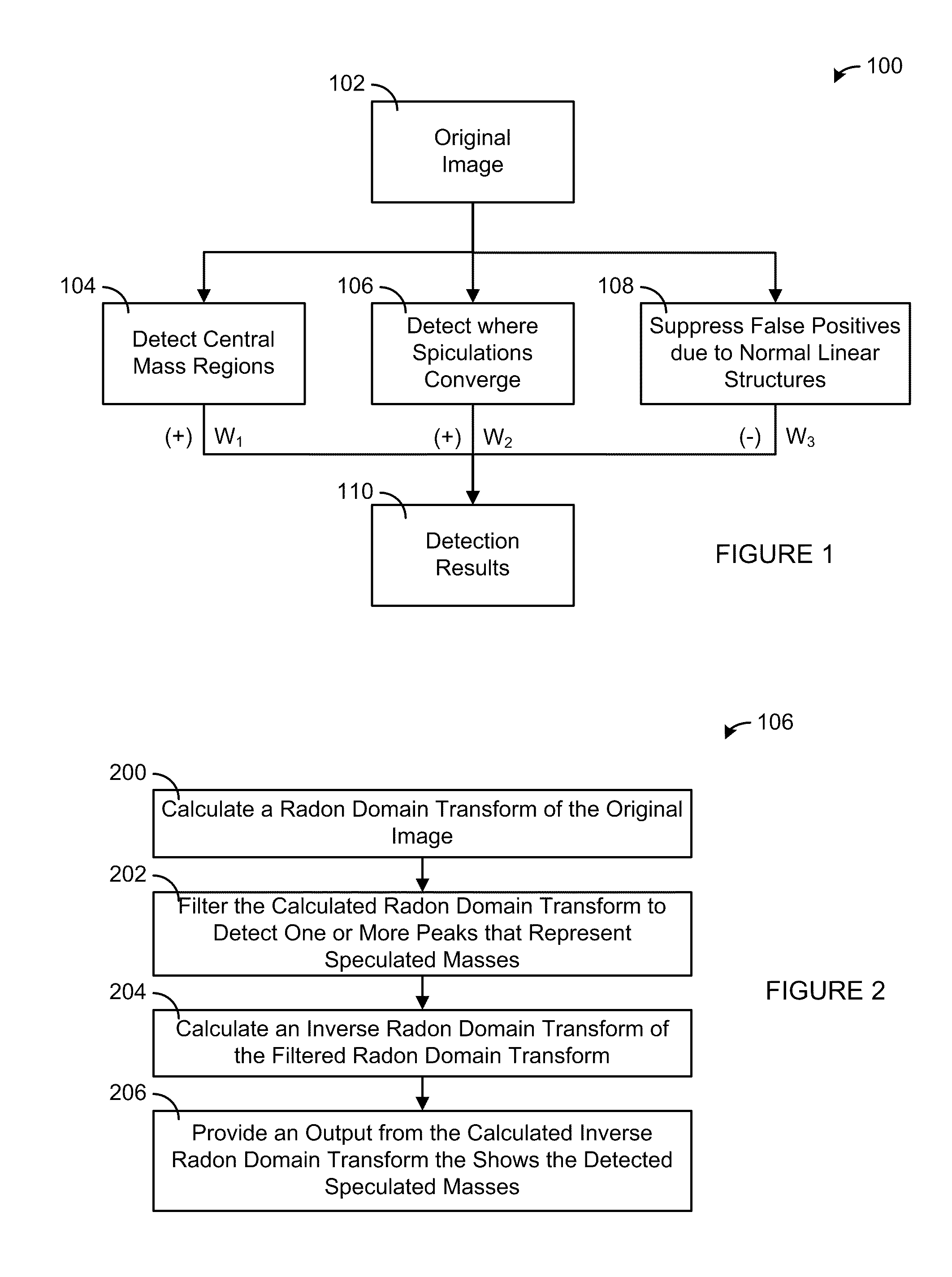
[0019]The step of performing spiculation filtration may further include the steps of computing a radon transformation of the image thereby producing a radon domain; filtering the radon domain using Spiculated Lesion Filters thereby forming patterns of line structures; and applying an inverse radon transformation to the patterns of line structures thereby obtaining an enhanced image with identifiable lines.
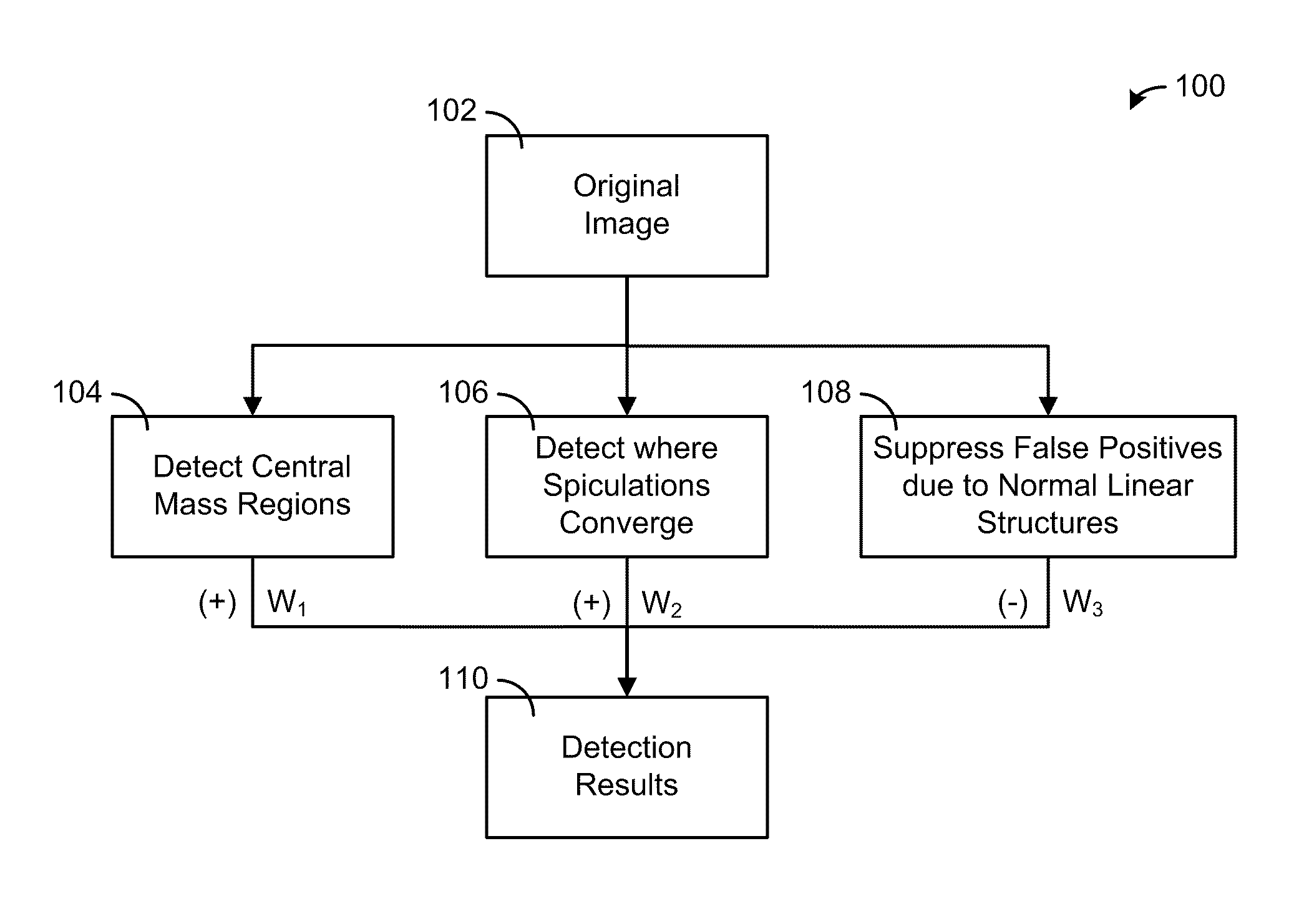
[0022]In yet another embodiment, the present invention provides an apparatus having a processor, a memory communicably coupled to the processor, and one or more communication interfaces communicably coupled to the processor. The processor receives an image from the communication interface or the memory, filters the received image using one or more Gaussian filters to detect one or more central mass regions, filters the received image using one or more spiculated lesion filters to detect where the one or more spiculated masses converge, filters the received image using one or more Difference-of-Gaussian filters to suppress one or more linear structures, creates an enhanced image showing the detected spiculated masses by combining an output from all of the filtering steps, and provides the enhanced image to the memory or the communication interface.

Problems solved by technology

Masses with spiculated margins carry a much higher risk of malignancy than other types of masses or calcifications.
However, mammography is not perfect.
Detection of suspicious abnormalities is a repetitive and fatiguing task.
Liu and Delp noted that, in general, it is difficult to estimate the size of the neighborhood that should be used to compute the local features of spiculated masses.
Small masses may be missed if the neighborhood is too large and parts of large masses may be missed if the neighborhood is too small.
However, all of these references lack data on physical properties of spiculated masses and the old computer-aided algorithm generates many false positive detections.

Method used


Image

  • Method and apparatus for detecting spiculated masses in mammography
  • Method and apparatus for detecting spiculated masses in mammography
  • Method and apparatus for detecting spiculated masses in mammography

Examples


Embodiment Construction

[0040]While the making and using of various embodiments of the present invention are discussed in detail below, it should be appreciated that the present invention provides many applicable inventive concepts that can be embodied in a wide variety of specific contexts. The specific embodiments discussed herein are merely illustrative of specific ways to make and use the invention and do not delimit the scope of the invention.

[0041]To facilitate the understanding of this invention, a number of terms are defined below. Terms defined herein have meanings as commonly understood by a person of ordinary skill in the areas relevant to the present invention. Terms such as “a”, “an” and “the” are not intended to refer to only a singular entity, but include the general class of which a specific example may be used for illustration. The terminology herein is used to describe specific embodiments of the invention, but their usage does not delimit the invention, except as outlined in the claims.

[...


PUM


Abstract

A method and apparatus detects one or more spiculated masses in an image using a processor. The image is received in the processor. The received image is filtered using one or more Gaussian filters to detect one or more central mass regions. The received image is also filtered using one or more spiculated lesion filters to detect where the one or more spiculated masses converge. In addition, the received image is filtered using one or more Difference-of-Gaussian filters to suppress one or more linear structures. An enhanced image showing the detected spiculated masses is created by combining an output from all of the filtering steps. The enhanced image is then provided to an output of the processor.

Description

CROSS-REFERENCE TO RELATED APPLICATIONS[0001]This patent application claims priority to U.S. provisional patent application Ser. No. 61 / 049,326, filed on Apr. 30, 2008 and entitled “Model-Based Framework for the Detection of Spiculated Masses on Mammography,” which is hereby incorporated by reference in its entirety.STATEMENT OF FEDERALLY FUNDED RESEARCH[0002]This invention was made with U.S. Government support under Contract No. W81XWH-04-1-0406 awarded by the U.S. Army Medical Research and Materiel Command. The government has certain rights in this invention.TECHNICAL FIELD OF THE INVENTION[0003]The present invention relates in general to the field of image processing and more particularly to the computer-aided detection of breast cancer.BACKGROUND OF THE INVENTION[0004]Without limiting the scope of the invention, its background is described in connection with computer-aided detection of breast cancer. The American Cancer Society estimates that 178,480 women will be diagnosed with...

Claims


Application Information

Patent Timeline
IPC IPC(8): G06K9/40G06V10/48
CPCG06K9/4633G06K2209/053G06T5/20G06T2207/30068G06T7/403G06T2207/10116G06T7/0012G06T7/44G06V10/48G06V2201/032
Inventor BOVIK, ALAN C.MARKEY, MIA K.SAMPAT, MEHUL
Owner BOARD OF RGT THE UNIV OF TEXAS SYST
Learn More