Method and device for selecting body model positions for SAR monitoring of a magnetic resonance transmit array

a magnetic resonance transmit array and body model technology, applied in the direction of magnetic measurement, geological measurement, reradiation, etc., can solve the problems of high degree of complexity, inability to identify the local sar in an mr scanner, and inability to monitor individual selected potential “hotspots” in clinical practi

Inactive Publication Date: 2010-12-09
SIEMENS HEALTHCARE GMBH
View PDF8 Cites 15 Cited by
  • Summary
  • Abstract
  • Description
  • Claims
  • Application Information

AI Technical Summary

Benefits of technology

[0009]An object of the present invention is to provide a method to systematically select model poin...

Problems solved by technology

Identification of the local SAR in an MR scanner has not previously been possible by means of measurement in clinical practice.
Comprehensive monitoring in the local SAR thus would involve a high degree of complexity, but ...

Method used


Image

  • Method and device for selecting body model positions for SAR monitoring of a magnetic resonance transmit array
  • Method and device for selecting body model positions for SAR monitoring of a magnetic resonance transmit array
  • Method and device for selecting body model positions for SAR monitoring of a magnetic resonance transmit array

Examples


Embodiment Construction

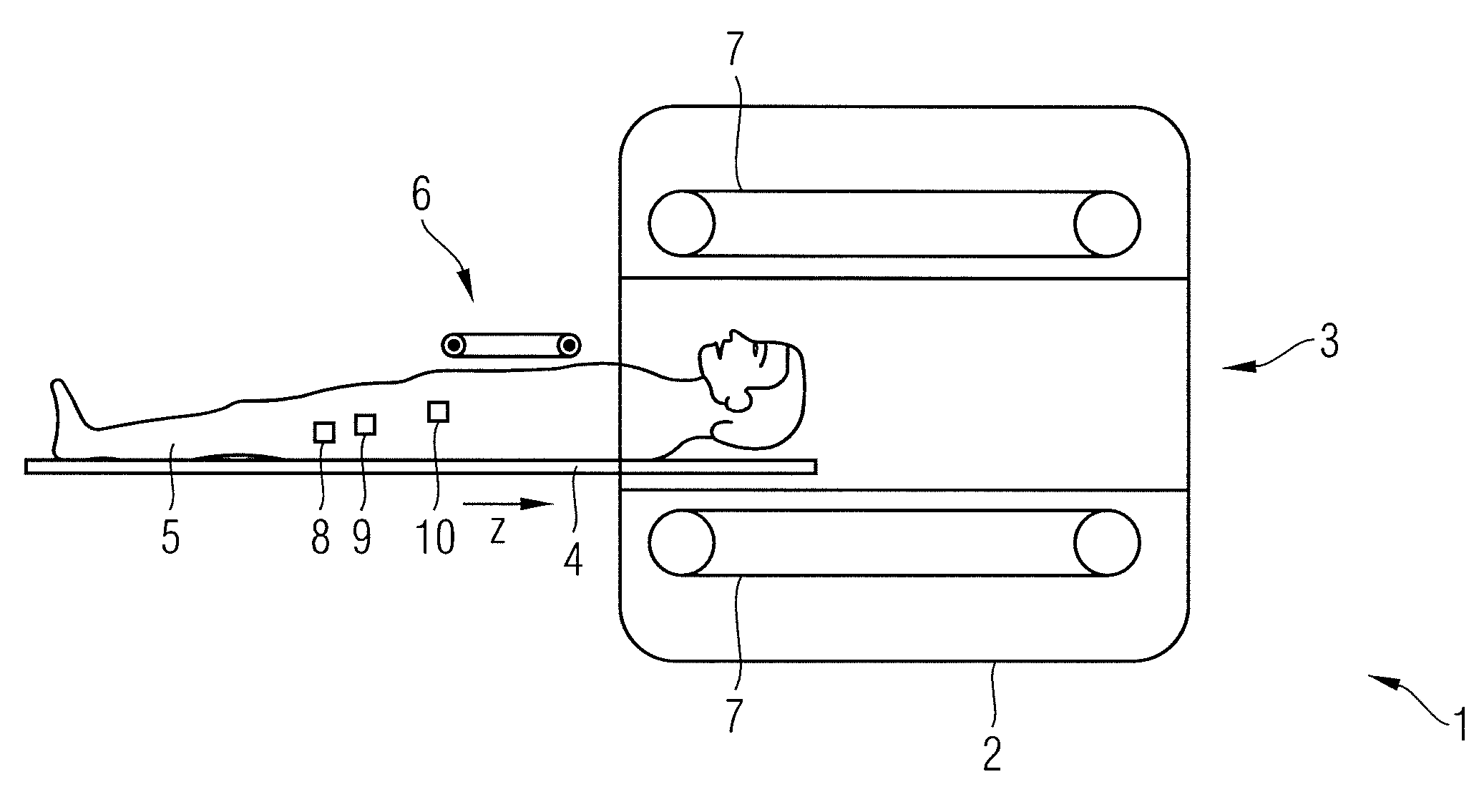
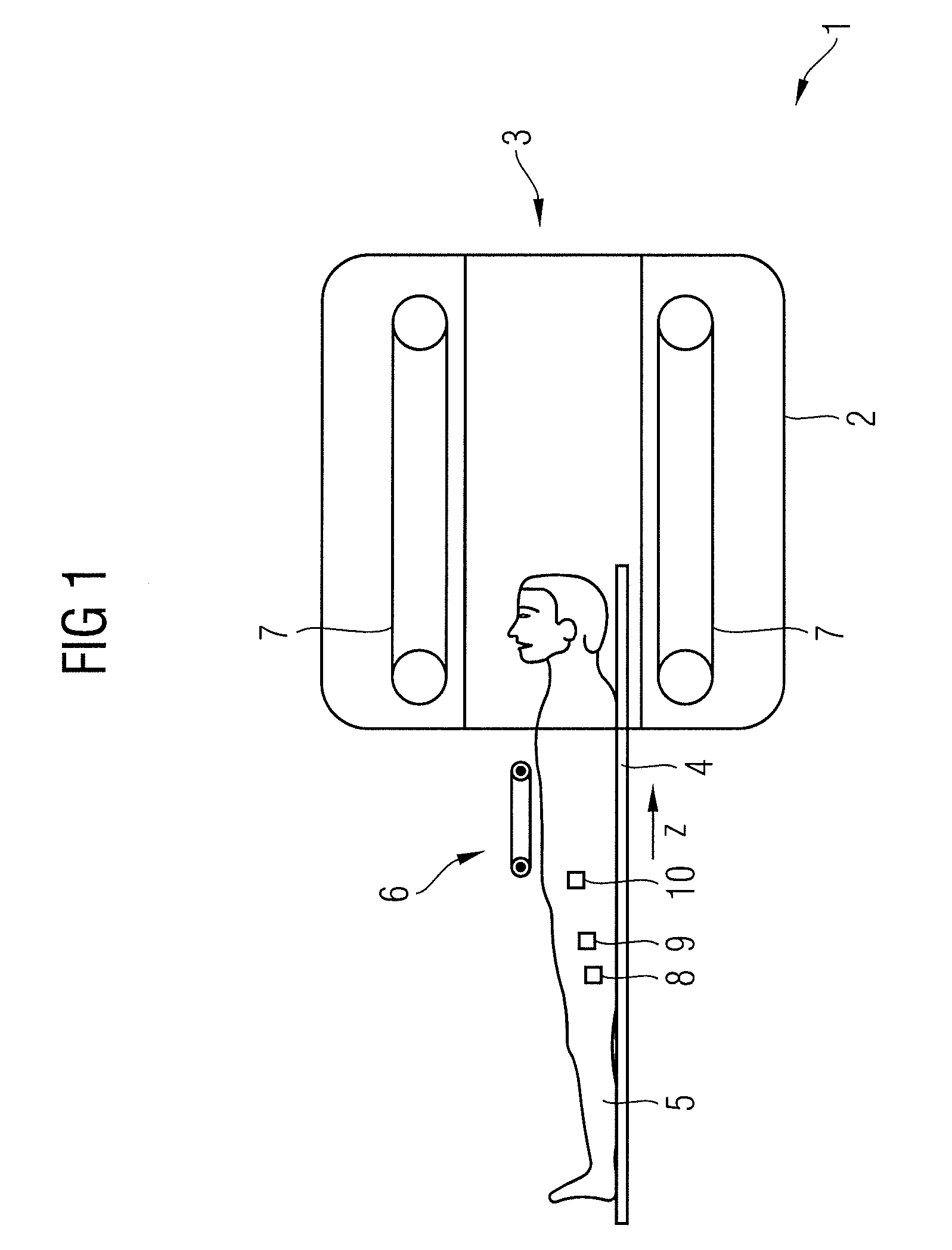
[0030]FIG. 1 shows a magnetic resonance apparatus 1 with a whole-body coil 2 and a tube-shaped space 3 into which a patient bed 4 with, for example, a patient 5 and a local coil 6 can be driven in order to generate exposures of the patient 5. Here a local coil with which good exposures are enabled in a local region is placed on the patient. The maximum SAR in the patient 5 should be monitored; for this multiple points 8, 9, 10 (also called model points 8, 9, 10 in the following) in the patient 5 should be determined at which absolute or relative maximum power density is realized (thus a greater power density than at other points in the patient). The point at which the highest power density occurs is called the primary model point voxel 8 in the following; points at which lower power density occurs than at the primary model point voxel 8 (but more than is expected elsewhere in the patient) are called auxiliary model point voxels 9, 10 (voxels are small volumes, for example small cubo...


PUM


Abstract

In a method and a device for specific absorption rate monitoring in a magnetic resonance system wherein multiple transmit coils are independently charged with respective currents, a primary model point voxel and at least one auxiliary model point voxel are automatically selected from among multiple voxels that model a modeled examination subject. The primary model point voxel is that voxel in which an absolute maximum of a total field variable occurs that is produced by the respective electrical fields emitted by the transmit coils. The at least one auxiliary model point voxel is that voxel in which a relative maximum of the variable occurs. The primary model point voxel and the at least one auxiliary model point voxel are stored, and specific absorption rate monitoring of an actual examination subject in the magnetic resonance system is implemented during the acquisition of magnetic resonance data in respective voxels of the actual examination subject corresponding to the stored primary model point voxel and the stored at least one auxiliary model point voxel.

Description

BACKGROUND OF THE INVENTION[0001]1. Field of the Invention[0002]The present invention concerns methods and devices for monitoring an examination subject in a magnetic resonance apparatus.[0003]2. Description of the Prior Art[0004]Identification of the local SAR in an MR scanner has not previously been possible by means of measurement in clinical practice. The only known and used technique is to make use of a theoretical simulation (model) that accounts for both the patient and the structure of the transmission antenna as an electrical model. In the operation of MR scanners with a transmission array and with RF pulses that can exhibit an arbitrary pulse shape for each array element (variation of amplitude and phase), a multitude of overlay possibilities result. Comprehensive monitoring in the local SAR thus would involve a high degree of complexity, but monitoring of the local SAR value is absolutely necessary for the safety of the patient and is required by corresponding regulations...

Claims


Application Information

Patent Timeline
IPC IPC(8): G01R33/44
CPCG01R33/5612G01R33/288
Inventor BRINKER, GERHARDDIEHL, DIRKGEBHARDT, MATTHIASNISTLER, JUERGENSCHNETTER, VOLKER
Owner SIEMENS HEALTHCARE GMBH
Why Eureka
Try Eureka