Process and apparatus for lung nodule segmentation in a chest radiograph

a chest radiograph and segmentation process technology, applied in the field of diagnostic medical imaging, can solve the problems of difficult segmentation, cumbersome medical doctors, and difficulty in automatic computerized detection of pulmonary nodules, and achieve the effect of fast and robust segmentation process

Inactive Publication Date: 2011-02-24
CARESTREAM HEALTH INC
View PDF5 Cites 3 Cited by
  • Summary
  • Abstract
  • Description
  • Claims
  • Application Information

AI Technical Summary

Benefits of technology

[0009]Another objective of this invention is to provide a fast and robust segmentation process by incorporating the visual features of pulmonary nodules, e.g., shape, density and texture of nodules.

Problems solved by technology

However the automatically computerized detection for a pulmonary nodule is very often faced with many difficulties due to the existence of the complicated anatomical structures in the chest radiograph.
This is because in many situations the nodules themselves do not have clear edges, and their size and shape may vary from case to case and thus incur cumbersomeness to the medical doctors.
In addition, many suspicious nodules may be sometimes superimposed on other organisms or anatomical structures, e.g., on ribs, and are thus difficult to be segmented.
Many methods have been thus far developed to have limitations to undesirable segmentation of nodules.
However this method may often lead to an over-segmentation with a very high computational cost and the clustering and merging of over-segmented sub-regions may incur another problem, e.g., the irregular shape of segmented contours.
However this latter method needs to predetermine a stopping threshold and is hence lack of robustness.

Method used


Image

  • Process and apparatus for lung nodule segmentation in a chest radiograph
  • Process and apparatus for lung nodule segmentation in a chest radiograph
  • Process and apparatus for lung nodule segmentation in a chest radiograph

Examples


Embodiment Construction

[0021]Generally, image segmentation is a process for partitioning a digital image into disjoint sets of connected pixels, one of which corresponds to the background and the remainders to the objects in the image which in the medical diagnosis may suggest an anatomical structure. Image segmentation can be approached as the process for either assigning pixels to the objects, or finding boundaries between the objects or between the objects and the background.

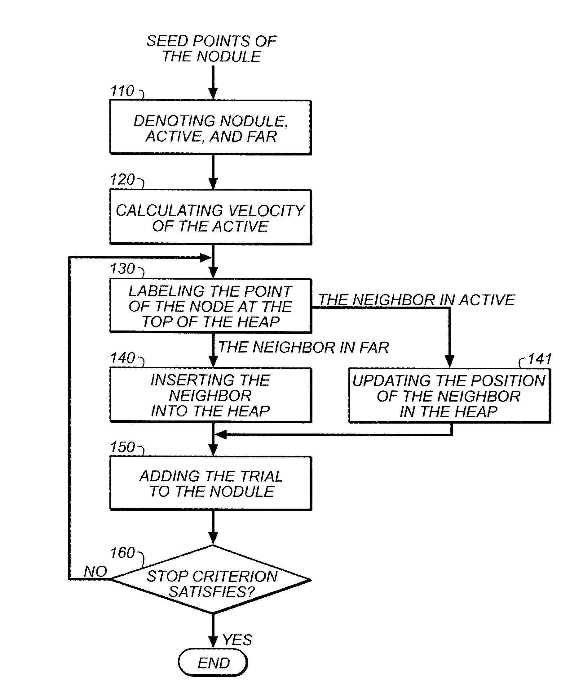
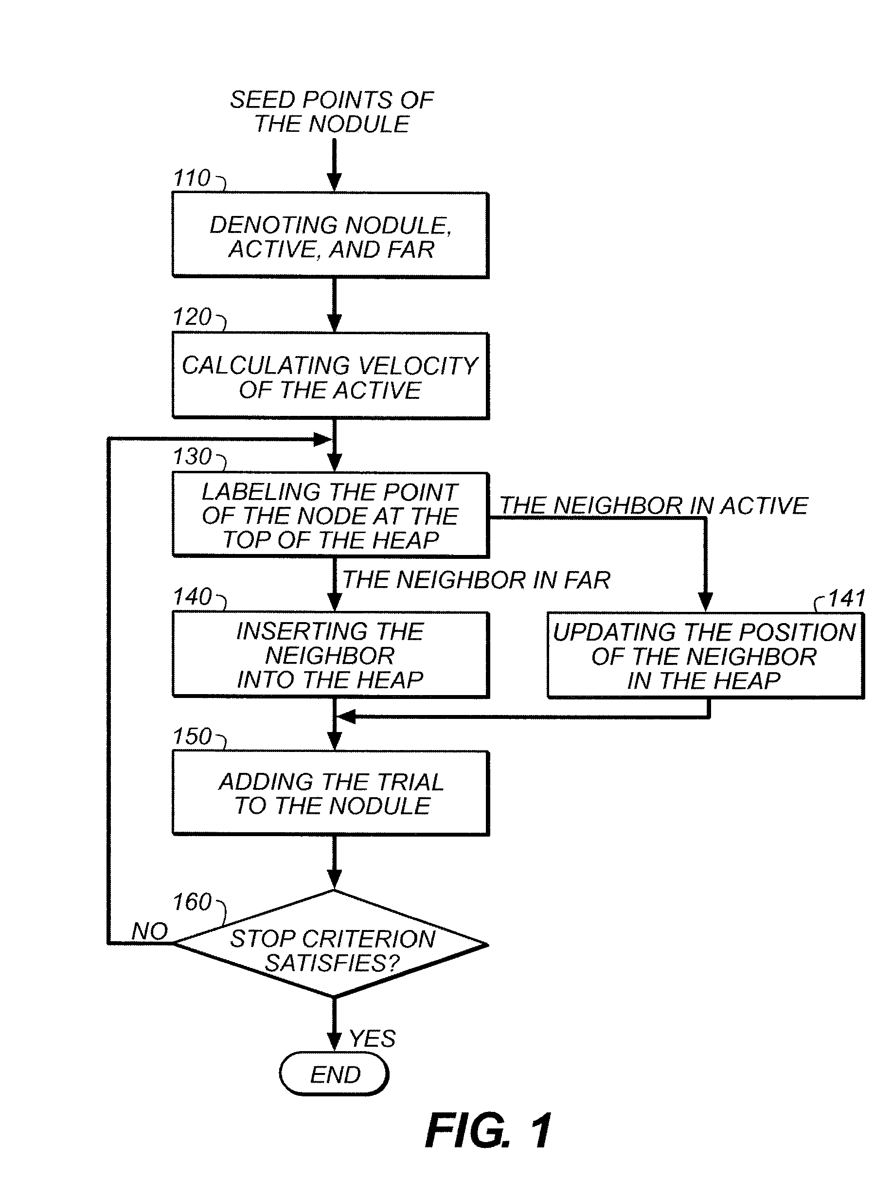
[0022]The non-restrictive illustrative embodiments of the present invention relate to a process and an apparatus for segmenting the nodule in a chest radiograph, in other words, a process and an apparatus for finding the boundary between a nodule and the background.

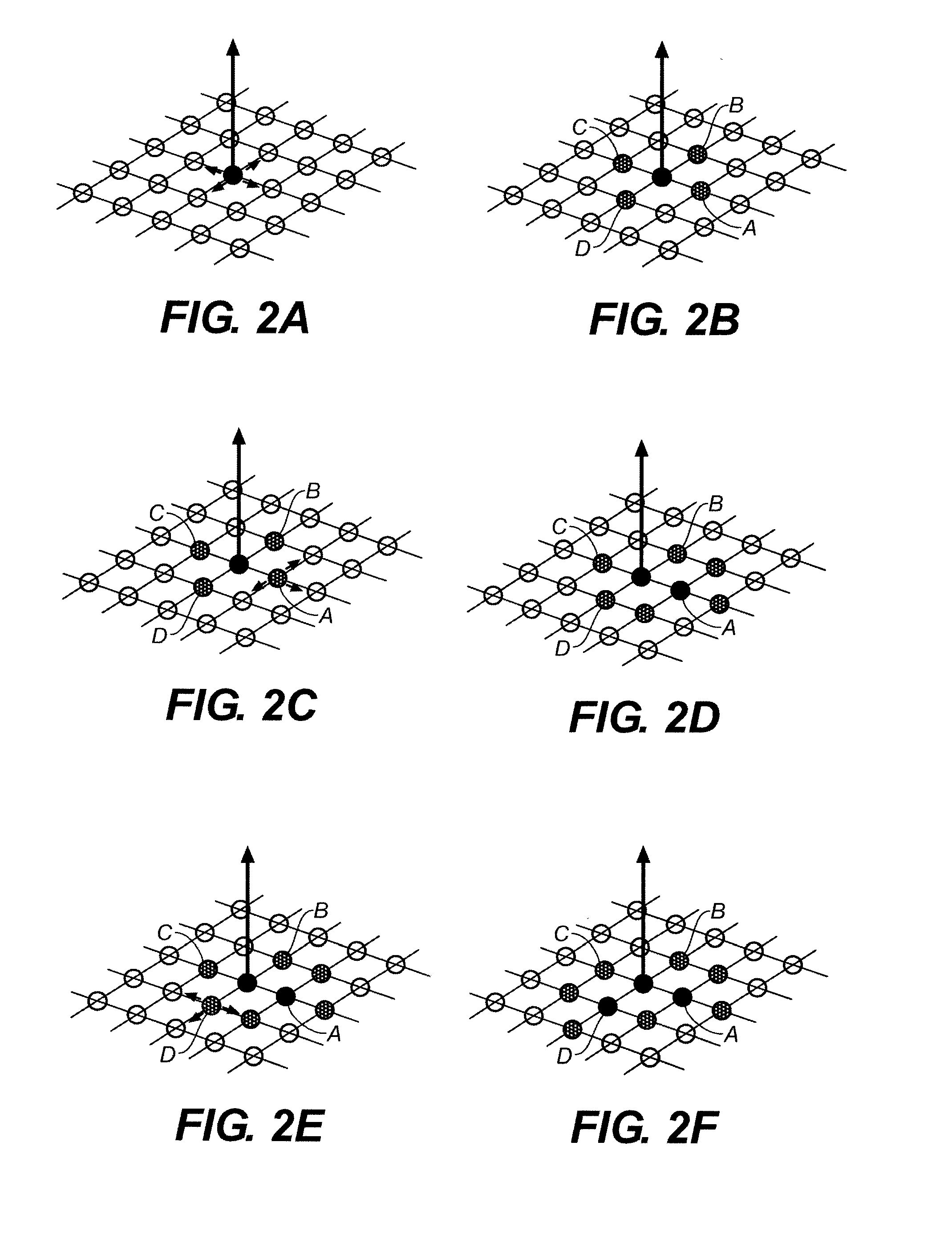
[0023]The fast marching (FM) method adopted in the present invention is illustrated in FIG. 3. Suppose there is an area which for simplicity of discussion is schematically depicted as an initial circle in FIG. 3. This initial circle schematically represents the initial s...


PUM


Abstract

The present invention has disclosed a method and apparatus for lung nodule segmentation in a chest radiograph. The method comprises preprocessing the chest radiograph and propagating the segmentation in the image based on the fast marching method. The method further includes design of velocity function. With the present invention, a fast and robust segmentation can be achieved.

Description

TECHNICAL FIELD[0001]The present application relates to diagnostic medical imaging and in particularly to the Computer Aided Detection (CAD) of pulmonary nodules in a chest radiograph.BACKGROUND OF THE INVENTION[0002]Lung cancer is one of the most common cancers. In 2007, lung cancer accounts for approximately 15% of all cancer diagnoses and 29% of all cancer deaths. It ranks to be the second mostly diagnosed cancers and the first cause of cancer deaths each year in our human being. Smoking has been substantiated to be a direct reason that leads to lung cancers, in addition to some other factors such as exposure to asbestos, radon, environmental deterioration, or secondhand smoking. In this regard, early diagnosis has been evidenced to be quite important.[0003]For an early and a correct diagnosis of the lung cancer, chest radiographs are a very commonly adopted measure by the medical doctors to discover the lung cancer which, in a chest radiograph, is in many situations shown to be ...

Claims


Application Information

Patent Timeline
Patent Type & Authority Applications(United States)
IPC IPC(8): G06K9/00
CPCG06T7/0081G06T2207/30064G06T2207/10116G06T7/0097G06T7/11G06T7/174
Inventor LIU, CHAOTAN, YONGQIANGLI, LIHONGXU, MANTAOZHANG, JIWU
Owner CARESTREAM HEALTH INC
Why Eureka
Try Eureka